Enzyme-responsive peptide dendron nanoassemblies for targeting and eliminating intracellular drug-resistant bacteria - Nature
High confidence: full text extraction produced 114571 characters.
Abstract
Certain intracellular pathogens can reside within cells to evade host immune defenses and exhibit high tolerance to antibiotics. Current treatments for intracellular bacterial infections are limited by poor cellular penetration, inadequate targeting of infected sites, and inefficacy against drug-resistant bacteria. Here, peptide dendron nanoassemblies comprising self-assembling regions, cell-penetrating motifs, enzyme-responsive sequences, and targeting ligands are developed to eliminate intracellular drug-resistant bacteria. The peptide dendrons self-assemble into nanoparticles that bind to membrane integrins via targeting sequence. The receptor-ligand interaction triggers a structural transformation into nanofibers for prolonged retention on the membrane surfaced of infected cells. Subsequently, they transform to nanoparticles upon tailored by bacteria-secreted enzyme, facilitating cellular uptake to target and eradicate intracellular drug-resistant bacteria. Self-assembling peptide dendrons further enhance macrophage resistance to infection-induced damage by modulating ferroptosis. The in vivo efficacy of peptide dendron nanoassemblies in removing intracellular drug-resistant bacteria is demonstrated in male mice and piglet infection models. This study provides a promising therapeutic strategy for treating intracellular drug-resistant infections.
Introduction
Intracellular bacterial infections pose a significant challenge in the medical field, particularly in the effective treatment of bacterial diseases due to the unique ability of certain pathogens to invade and persist within host cells1. Pathogens such as Mycobacterium tuberculosis, Salmonella enterica, along with the ESKAPE “superbugs” including Staphylococcus aureus listed by the World Health Organization (WHO), possess the ability to survive and replicate within host cells and have evolved diverse and sophisticated evasion strategies to escape host immune responses2,3. Even though immune cells such as macrophages serve as the primary line of defense against and clearance of pathogens4, these cunning pathogens not only undermine the clearance mechanisms but also hijack macrophages as protective niches, allowing them to resist the bactericidal effects of high dose of antibiotics5. The current standard treatment for diseases associated with intracellular bacterial infections relies on prolonged and intensive antibiotic therapy6. However, poor antibiotic penetration, limited intracellular concentrations, and the complexity of the intracellular environment significantly constrain antimicrobial efficacy, making clinical treatment failures and frequent relapses increasingly common6. The emergence of multidrug-resistant (MDR) bacteria further complicates treatment, as it not only fails to prevent the invasion of host cells by extracellular bacteria but also makes it significantly more difficult to eliminate MDR pathogens residing within host cells. Collectively, there is an urgent need to develop strategies capable of effectively penetrating cells and targeting intracellular drug-resistant bacteria for eradication, without posing a risk of resistance development.
Peptide-based antimicrobial materials with cell-penetrating properties have garnered significant attention as effective antibiotic candidates for combating intracellular pathogens7,8. The inherent structural flexibility, chemical modifiability, and programmable self-assembly characteristics of peptide enable precise engineering of multifunctional biomaterials with tailored properties, notably including efficient cellular penetration9,10. The unique membrane-disrupting mechanisms of peptides, which rely on physical interactions rather than specific molecular targets, significantly reduce the risk of resistance development9. These advantages establish peptide-based antimicrobial materials as a promising platform to overcome the limitations of traditional antibiotics in treating intracellular bacterial infections. As research into peptide supramolecular self-assembly deepens, peptides can be designed and assembled into various forms of peptide-based nanomaterials. The structurally well-defined peptide assembly has better performance than the peptide itself, including enhanced stability in biological environments, improved cellular uptake efficiency, and the ability to form specific morphologies and dimensions through controlled self-assembly, thereby adapting to diverse application requirements11,12. However, a contradiction exists between size and biological function in cell-penetrating nanomaterials. Small-sized peptide nanoparticles can easily penetrate cell membranes but have short retention times at infection sites and are prone to rapid clearance by the body. In contrast, large-sized peptide nanofibers exhibit prolonged tissue retention but have poor penetration efficiency13,14. Previous studies have shown that filamentous nanofibers with a length of approximately 600 nm exhibiting prolonged circulation and retention compared with spherical nanoparticles, whereas small-sized spherical nanoparticles of about 20 nm displayed superior penetration capability15. Similarly, another study reported that large network-like nanofibers provide enhanced retention, while their transformation into smaller nanospheres of around 25 nm facilitated efficient cellular penetration16. Therefore, manipulating the size transformation of peptide self-assembly structures is crucial to achieving effective therapeutic outcomes. Current design strategies for peptide nanostructure transformation predominantly involve receptor-ligand recognition systems (e.g., cellular membrane receptors, pathogen-specific membrane components) and stimulus-responsive adaptations to infection or physiological environment alterations (e.g., pH, enzymes, redox conditions)11,17,18,19. While these approaches have demonstrated efficacy in managing superficial infections and localized tissue colonization by planktonic bacterial, they have not been effectively implemented to address recalcitrant intracellular bacteria.
Another dilemma arises from the lack of targeting in cell-penetrating peptide nanomaterials, which manifests as two critical challenges: reduced therapeutic efficiency and potential off-target cytotoxicity. This non-targeted distribution pattern significantly diminishes the local concentration of antimicrobial agents at infection sites while potentially inducing adverse effects in non-target cells. Furthermore, the widespread cellular uptake of peptide-based nanomaterials may trigger unintended biological responses, including immune activation or disruption of normal cellular functions. The altered microenvironment at bacterial infection sites presents a promising opportunity for the development of intelligent targeted antibacterial strategies. Hence, addressing the poor targeting specificity and the size paradox in cell-penetrating peptide-based nanomaterials is critically important and urgently required.
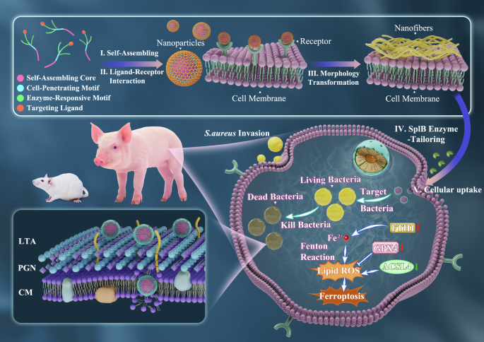
In this work, we design a multifunctional chimeric peptide nanoassembly integrating with cell-penetrating, structural transformation, and microenvironment-responsive properties to achieve targeting of infected macrophages and precise clearance of intracellular MRSA. The modular design of peptide nanomaterials consists of four distinct functional units: (1) the N-terminal myristic alkyl chain (C-14) is strongly hydrophobic to force the self-assembly of the peptides into stable nanostructure20; (2) the proline-arginine-arginine-isoleucine (PRRI) motifs responsible for cell-penetrating are repeated or attached to the branched chains of the peptide backbone to perform as dendritic structures; (3) the enzyme cleavage fragment (tryptophan-glutamic acid-leucine-glutamine, WELQ) serves as the responsive motif, and the enzyme serine protease-like B enzyme proteins (SplB) secreted by MRSA specifically recognizes and cleaves the peptide bond following Gln in the sequence21,22; (4) the introduction of the tripeptide sequence (arginine-glycine-aspartic acid, RGD) at the C-terminus, which interacts with the αvβ3 integrin receptor on the membrane surface of the macrophage to increase adhesion to the macrophage and promote cellular uptake23,24. The chimeric peptides self-assemble into nanoparticles driven by hydrophobic interactions, which convert into nanofibers upon ligand-receptor interactions between integrins expressed on target cells and the RGD motif, enabling specific accumulation and prolonged retention at infection sites. Subsequently, the nanofibers are tailored by bacterial-secreted enzymes into small-sized nanoparticles, facilitating efficient cellular penetration and targeted delivery to intracellular MRSA. Self-assembling peptide nanoassemblies eliminate intracellular bacteria through a membrane-disruptive mechanism that minimizes the risk of resistance development, while protecting macrophages from damage induced by ferroptosis (Fig. 1), showing promise for treating intracellular drug-resistant bacterial infections.
Results and discussion
Design, synthesis, and characterization of self-assembling chimeric peptides
To precisely target macrophages infected by bacteria, we developed a ligand-receptor interaction and enzyme-responsive self-assembling peptide nanomaterial, leveraging the principles of peptide self-assembly and the structure-function relationship of antimicrobial biomaterials. Specifically, peptide molecules depend on C-14 alkyl chains to self-assemble ordered nanostructures driven by hydrophobic interactions25. Previous studies have reported a class of non-membrane-lytic antimicrobial peptides capable of penetrating cell membranes and inhibiting the translation process by targeting intracellular ribosomes26,27. These peptides such as PR-39 and bactenicin are characterized by their high content of proline (Pro) and arginine (Arg) residues with PRP repeats in the sequence. Increasing the Arg content with PRR motifs has been shown to improve cellular uptake efficiency, where the positively charge and guanidinium group of Arg are key determinants for membrane penetration28,29. In our design, a hydrophobic amino acid, isoleucine (Ile), was introduced to enhance the antimicrobial activity, resulting in the cell-penetrating repeat motif PRRI. Subsequently, we connected this motif to the peptide backbone by a branched chain to develop a peptide dendritic structure and compared it with linear peptide of the same amino acid composition, in order to better explore the relationship between the structure and the biological function of peptide nanomaterials. The WELQ sequence can be specifically recognized and cleaved by SplB, a protease secreted by MRSA, making the peptide with MRSA-targeting and enzyme-responsive properties22. Finally, the modification of the C-terminal RGD peptide served as a targeting motif allowing the self-assembling peptide to target the αvβ3 integrin receptor on macrophages through ligand-receptor recognition23,24, increasing peptide accumulation followed by facilitating efficient peptide uptake (Fig. 2a and Supplementary Fig. 1). The rationally designed branched peptide dendron (BCPR) and linear chain peptide (LCPR) were synthesized using solid-phase synthesis and purified by reverse-phase high-performance liquid chromatography (RP-HPLC). The synthesized peptides were determined by RP-HPLC and mass spectrometry (ESI-MS), which proved that the purity greater than 95% and the observed molecular weights were identical to the theoretical value (Supplementary Fig. 2 and 3a, b).
The critical aggregation concentration (CAC) of the peptide was first determined using the fluorescent dye 8-anilino-1-naphthalenesulfonic acid (ANS), which was hydrophobic-affinity, to characterize their self-assembly behavior30. As the concentration of the peptide increased, the fluorescence intensity of ANS showed a sharp enhancement, demonstrating that the peptides self-assembled to form a hydrophobic core region (Fig. 2b–d). Two lines were fitted through the fluorescence intensity of the peptides at 480 nm, and the intersection of the lines identified as CAC. The CACs for BCPR and LCPR were calculated to be 17.8 μM and 17.4 μM from Fig. 2e, f, respectively. It was shown that the concentration required for the self-assembly of BCPR and LCPR into nanostructures did not differ much, which was due to the fact that their hydrophobic compositions were the same and that the process was controlled by the hydrophobic alkyl chains9. To further validate the formation of the peptide nanostructures and characterize the size, transmission electron microscopy (TEM) and dynamic light scattering (DLS) experiments were performed. Both BCPR and LCPR were observed to self-assemble into nanoparticles and were uniformly dispersed with sizes of 10.02 ± 2.79 nm and 9.25 ± 1.38 nm under TEM, respectively (Fig. 2h, i). Subsequently, the hydrodynamic diameters of the peptide nanoparticles were measured by DLS, and the sizes were consistent with those observed by TEM (Fig. 2j and Supplementary Fig. 3c). The zeta potentials of BCPR and LCPR were 17.07 ± 1.25 mV and 10.21 ± 1.60 mV, respectively (Fig. 2k), indicating that the surfaces of the self-assembling peptide nanomaterials are positively charged. The structure of branched and linear chains affected the surface charge of the peptide nanoparticles, and even though they contained the same number of positive charge amino acids (Arg), the branched structure contributed to an increase in the surface positive charge density of the peptide nanomaterials, which facilitated interaction with the cell membrane and promoted cellular uptake31,32.
Next, the secondary structure of the peptides was identified by circular dichroism (CD). As shown in Fig. 2g, the branched and linear structures did not influence the secondary structure of the peptides, and both the BCPR and LCPR exhibited a random coil conformation, as characterized by a negative peak at about 200 nm. This was due to the Pro residue presented in the sequence, which had a rigid structure that interfered with the formation of hydrogen bonds between amino acids, thereby preventing the formation of α-helical and β-sheet conformations33. This was consistent with the structural properties of Pro-rich peptides, which exhibited low cytotoxicity, thus allowing the peptides to penetrate cells without membrane damage10,34,35. We designed self-assembling peptides in response to proteases secreted by MRSA and therefore revealed their enzyme responsiveness by HPLC. For the purpose of verification, we additionally synthesized the sequences of BCPR and LCPR after they were enzymatically cleaved, named BCP (C14-K(IRRP)K(IRRP)PRRIWELQ) and LCP (C14-PRRIPRRIPRRIWELQ), respectively (Supplementary Fig. 4 and 5). The HPLC results showed retention times of approximately 12 and 12.5 min for BCPR and BCP, respectively, and 15.3 and 16 min for LCPR and LCP, respectively (Fig.2l, m). When interacted with the SplB protease for 12 h, the retention times of BCPR and LCPR were changed to coincide with the sequences after cleavage, indicating the enzyme responsiveness of peptides.
Biological properties of self-assembling chimeric peptides
Following the structural characterization of the designed peptides, their antimicrobial capacity was evaluated, and the structure-function relationship was further explored. The results of minimum inhibitory concentrations (MICs) and minimum bactericidal concentrations (MBCs) against MRSA showed that BCPR and LCPR exhibited favorable antibacterial activity both before and after enzymatic reaction, and the effects of peptides after interaction with enzyme were in accordance with those of synthesized BCP and LCP, which further confirming the enzyme-responsiveness of the peptides (Fig. 3a). To more comprehensively investigate the antibacterial potential of the self-assembling chimeric peptides, we additionally selected several strains of methicillin-sensitive Staphylococcus aureus (MSSA) and determined the MICs and MBCs of the peptides against them. The results demonstrated that chimeric peptides exhibited potent antibacterial efficacy, capable of killing 99.99% of the bacteria at low concentrations. In addition, BCPR exhibited superior antibacterial activity compared to LCPR, which might be attributed to the branched structure increasing the positive charge density on the peptide surface, thereby enhancing the interaction with the bacterial membrane and improving its antibacterial activity. This observation was consistent with the conclusions derived from the zeta potential measurements. After confirming that the peptides effectively targeted extracellular bacteria, it was essential to investigate whether the peptides exert any adverse effects on eukaryotic cells, including red blood cells, which was a prerequisite for determining the potential application of these peptides as intracellular antimicrobial agents. The results of hemolysis assays showed that BCPR and LCPR maintained hemolysis levels below 10% even at a high concentration of 128 μM (Fig. 3b). In contrast, the peptide antibiotic polymyxin B exceeded 10% hemolysis at 32 μM. To evaluate the cytotoxicity of the peptide toward mammalian cells, we performed combined MTT assays and lactate dehydrogenase (LDH) assays in RAW264.7 cells, primary porcine alveolar macrophages, and HEK293T cells. The cell viability above 80% was defined as the threshold for cytotoxicity36. The results showed that at a concentration of 128 μM, the peptide exhibited mild toxicity toward RAW264.7 cells, while no cytotoxicity was observed in primary alveolar macrophages or HEK293T cells (Fig. 3c–e and Supplementary Fig. 6). Differences in toxicity among cell lines may be associated with variations in membrane receptor distribution, membrane lipid composition, endocytosis, and metabolic status.
Subsequently, their bactericidal activities against MRSA within macrophages were assessed at non-toxic concentrations. The peptides effectively eliminated intracellular MRSA, as shown in Fig. 3f, h. At a concentration of 32 μM, BCPR and LCPR achieved intracellular MRSA clearance rates of 99.9% and 89.7%, respectively. Furthermore, BCPR not only exhibited significantly greater efficacy against intracellular bacteria compared to LCPR but also than that of the antibiotic vancomycin, which served as the primary treatment for the drug-resistant Gram-positive bacteria37,38. Next, we used confocal microscopy with Z-stack imaging to observe the state of intracellular MRSA after treatment with the peptides and vancomycin. We constructed a GFP-labeled MRSA ATCC43300 strain to establish an intracellular infection model and stained the cell cytoskeleton with rhodamine-labeled phalloidin, enabling clear visualization of the intracellular localization of the bacteria (Fig. 3i). Multiple clusters of MRSA were visible inside the untreated cells, when peptides or vancomycin were treated, the number of bacteria was reduced with a small amount of green fluorescence, particularly in the BCPR-treated cells. This fluorescence reduction was attributed to the release and degradation of GFP following bacterial death, indicating that BCPR effectively kills intracellular MRSA, which was similar to the findings from the plate colony counting assay (Fig. 3h). The bactericidal kinetics against intracellular MRSA revealed that BCPR exhibited a faster killing rate compared to LCPR. At a concentration of 32 μM, BCPR achieved complete bacterial clearance within 2 h, whereas at 16 μM, it required 6 h (Fig. 3g). These findings indicated that BCPR displayed more rapid bactericidal activity at concentrations exceeding its CAC, suggesting that the formation of nanostructures enhanced its efficacy against intracellular bacteria. Furthermore, the above results collectively indicated that BCPR exhibited stronger effects against extracellular and intracellular MRSA than LCPR. BCPR effectively eliminated intracellular bacteria at 16 μM, while it showed no evident cytotoxicity toward mammalian cells even at concentrations up to 64 μM and 128 μM, suggesting an acceptable therapeutic window. Therefore, we will treat it as a target peptide for subsequent studies. Under TEM, intact MRSA with full content was observed inside the infected cells (Fig. 3j). When the cells were treated with 16 μM of BCPR, a change in the morphology of MRSA could be observed, with the appearance of cavities inside the bacteria. When the concentration was increased to 32 μM, intracellular MRSA exhibited an obvious disrupted structure. Given that biofilm formation was also a key strategy by which bacteria evaded antibiotics and immune responses, thereby promoting the development of antimicrobial resistance39,40, we further evaluated the antibiofilm activity of BCPR. The bacterial viability within biofilms was determined using the MTT assay. The results showed that a significant bactericidal effect was observed at a concentration of 8 μM, and the bacterial mortality exceeded 80% at 32 μM (Supplementary Fig. 7a). The ability of peptide to disrupt mature biofilms was assessed using crystal violet staining. The results demonstrated that the BCPR could effectively eliminate pre-formed mature biofilms (Supplementary Fig. 7b). These findings suggested that the self-assembling peptide BCPR had the potential to combat biofilms, which might be attributed to its strong positive charge and penetrating capacity that facilitated its adsorption onto the biofilm surface and penetration into the biofilm matrix. Previous studies have shown that CPPs enhance biofilm penetration and clearance efficacy41,42.
Membrane receptor-mediated structural transformation and macrophage penetration of self-assembling peptides dendrons targeting intracellular MRSA
The RGD sequence is currently the most widely used and effective modification for peptide-based biomaterials, stimulating the targeting and adhesion of biomaterials to cells, and subsequently promoting cellular internalization23,43,44. Therefore, we introduced RGD modifications in peptide design to enhance the adhesion and internalization of self-assembling peptides to target cells. However, there are various studies that have reported that RGD acts as a targeting ligand that specifically binds to integrin αVβ3 on the cell membrane surface, and subsequently triggers the in situ self-assembly of peptides into nanofibers or the transformation of nanoparticles to nanofibers via receptor-ligand interactions45,46. In view of this, we first investigated whether the self-assembling peptide dendrons were also capable of structural transformation through interaction with integrin αVβ3. Previous findings have demonstrated that peptide dendron monomers can self-assemble into nanoparticles in aqueous solution (Fig. 4a). Interestingly, when the peptides were incubated with integrin αVβ3, the nanostructural morphology of the peptides was changed and nanofibers were observed under TEM (Fig. 4b). Following this, we examined the variation in the size of the peptide nanoassemblies upon their interaction with integrins using DLS. As shown in Fig. 4c, before the interaction with the integrin αVβ3 protein, the diameter of BCPR was approximately 10 nm. With the continuous increase in the ratios of peptide to integrin αVβ3 protein, the diameter changeed significantly, and when the protein-to-peptide ratio reaches 1:500, large-sized nanofibers could be clearly detected. These results demonstrated that peptide nanoparticles can transform into nanofiber networks through ligand-receptor interactions after binding with integrin αvβ3. Compared to small-sized nanoparticles, large-sized nanofibers prolong blood circulation time and can retain and act at the site of infection for a long time47. The induction of structural transformations by receptor-ligand interactions has been widely confirmed in several studies48,49. The self-assembly process could be induced by mechanical forces generated by intracellular actin filaments, which arose from the binding of protein ligands to cell surface receptors, and such forces were capable of triggering conformational changes or intermolecular rearrangements that promoted structural transformations of nanoparticles11,50.
The binding of peptides to membrane receptors may contribute to cellular internalization. Consequently, we assessed the ability of peptides to penetrate cells. We synthesized 5-TAMRA-labeled peptide dendron with red fluorescence to quantify their entry into the cells and to localize their position within the cells (Supplementary Fig. 8). Quantification of the peptides entering the cells by flow cytometry showed that 74.89% and 96.25% of the peptides were within the cells at peptide concentrations of 16 μM and 32 μM, respectively, demonstrating the high cell uptake of peptides (Fig. 4f). This enhanced penetration may be associated with the increased surface density of positive charges on the nanoparticles, as zeta potential measurements revealed a concentration-dependent increase in surface charge density (Supplementary Fig. 9). However, peptide dendrons penetration into cells was significantly inhibited when we pretreated RAW264.7 cells with RGD inhibitors. Flow cytometry results showed that the penetration efficiency of peptide dendrites was reduced from 86.77% to 55.12% and 58.74% after treatment with two RGD inhibitors, GRGDSP and RGDS, respectively (Fig. 4g). This result was in accordance with the confocal observation that when treated with the RGD inhibitors, intracellular red fluorescence was significantly attenuated compared to untreated RAW264.7 cells (Fig. 4e). The above results suggested that self-assembling peptide dendron nanoparticles could transform into nanofibers after interaction with receptors through the RGD ligand, thereby enhancing the accumulation and adhesion of the peptide on the macrophage cell membrane, which in turn facilitated the subsequent internalization process (Fig. 4h). We simulated the process in which the peptide bound to membrane receptors and was subsequently exposed to SplB. The size change of the peptide nanoassemblies after interaction with the receptor protein and undergoing enzymatic cleavage by SplB was detected by DLS. The self-assembling peptide initially increased in size after receptor interaction but subsequently decreased to approximately 5 nm nanoparticles following SplB treatment (Supplementary Fig. 10a). This result was consistent with the DLS measurements obtained directly from the cleaved peptide sequences, indicating that the conversion of peptide nanofibers into nanoparticles is specifically triggered by SplB (Supplementary Fig. 10b). To investigate the cellular uptake pathway of BCPR, we evaluated its penetration efficiency using flow cytometry under low-temperature conditions (4 °C) and in the presence of various inhibitors targeting distinct endocytic processes. The results showed that although low temperature suppressed cellular energy metabolism, BCPR still maintained a high uptake level (Supplementary Fig. 11), indicating that it primarily entered cells via an energy-independent direct translocation mechanism10. Moreover, none of the endocytic inhibitors exerted a significant effect on BCPR uptake, further supporting that its internalization was not endocytosis-dependent. The direct transduction of BCPR may be attributed to its small size (5 nm) following the enzyme response, which enables efficient membrane penetration and direct delivery into the cytoplasm, thereby exerting rapid biological effects. Confocal microscopy further revealed the intracellular distribution of BCPR (Fig. 4d). The red fluorescence of the peptide overlapped with the green fluorescence of lysosomal markers, and the intracellular fluorescence intensity increased with higher peptide concentrations. These findings suggest that after penetrating the cell, BCPR can reach lysosomes and interact with bacteria residing within, as most invading bacteria are initially enclosed and localized in lysosomes.
We then investigated the targeting of intracellular MRSA by peptide dendrons upon cellular internalization. TEM imaging revealed that after treatment with the peptide dendrons, distinct and uniformly distributed nanoparticles approximately 5 nm in size could be observed around both MRSA within the cells and intracellular MRSA released from lysed cells, whereas no nanoparticle was detected around intracellular MRSA in untreated cells (Fig. 5a and Supplementary Fig. 12). This result indicated that after the self-assembling peptide dendrons form nanofibers, they were followed cleaved by enzymes secreted by MRSA around the infected cells. This cleavage of the RGD sequence caused the peptides to transition from nanofibers back into nanoparticles, which were then internalized into the RAW264.7 cells to target intracellular MRSA. The large-sized nanofibers, while having the advantage of prolonged retention due to their higher specific surface area, faced the limitation of more difficult cell penetration and lower efficiency in internalization51. In contrast, small-sized nanoparticles can overcome these challenges, enabling efficient cellular penetration, which explained the high efficiency of peptide dendrons in penetrating macrophages13. Thereafter, intracellular GFP-labeled MRSA were observed to surrounded by the 5-TAMRA labeled BCPR under confocal microscopy (Fig. 5b). The killed MRSA no longer exhibited green fluorescence but was instead marked with red fluorescence by the peptides, suggesting that the BCPR entering the cell could target bacteria in RAW264.7 cells and thus exert antimicrobial effects. The morphological transformation induced by stimulus response facilitated the aggregation of peptide-based nanomaterials and their entry into target cells, thereby achieving enhanced functionality and therapeutic efficacy17.
Mechanism of self-assembling peptide dendron against drug-resistant bacteria
A major advantage of peptide-based nanomaterials in the field of antibacterial applications is their membrane-disrupting properties, a physical membrane-breaking mechanism that makes them less susceptible to drug resistance, providing a sustainable approach to combating infections52,53. The antibacterial action of peptide-based nanomaterials initially begins with electrostatic interactions between the peptides and the negatively charged components on the bacterial membrane surface54. The surface of MRSA was rich in negatively charged lipoteichoic acid (LTA), and the peptides we designed were cationic in nature, so, we first evaluated their binding affinity with LTA. As shown in the Fig. 5c, both BCPR and LCPR demonstrated a dose-dependent binding ability to LTA, as the concentration increased, their binding strength to LTA gradually enhanced. In addition, BCPR exhibited a stronger binding affinity for LTA compared to LCPR, which was attributed to the increase in surface positive charge density conferred by the branched structure, which led to more accumulation of BCPR on the surface of the bacterial membrane, thus enhancing the antimicrobial capacity of BCPR. As the peptides accumulate, they disrupt the bacterial membrane through the action of hydrophobic groups. To assess their effect on the membrane permeability of MRSA, we used the DISC3-5 membrane potential-sensitive probe. DISC3-5 aggregates within the phospholipid bilayer, causing self-quenching of the probe, which is released into solution to fluoresce continuously when the membrane potential is altered55. Both BCPR and LCPR treatment induced membrane depolarization in MRSA (Fig. 5d, e), and the fluorescence intensity enhanced with the increase of concentration. The fluorescence of the peptides increased dramatically within 120 s and then stabilized, indicating that they were able to penetrate the thick peptidoglycan layer of MRSA to reach the cytoplasmic membrane within a short period of time. Compared to LCPR, treatment with the same concentration of BCPR induced a more pronounced membrane depolarization response in MRSA, as evidenced by higher fluorescence intensity, indicating that BCPR caused greater damage to the MRSA membrane. The branched structure contributes to a more centralized contact of BCPR with the bacterial membrane surface, and besides that, it has been reported that the dendritic structure is also more likely to form pores that help the peptide to cross the bacterial membrane56,57.
Subsequently, we quantified self-assembling peptides treatment-induced bacterial death by flow cytometric analysis of the fluorescence intensity of propidium iodide (PI), a membrane-impermeable dye that can cross the membrane and bind to DNA to emit fluorescence only if the bacterial membrane is ruptured. Treatment of MRSA with BCPR at concentrations of 2 and 4 μM resulted in the death of 73.39% and 96.42% of MRSA, respectively, whereas treatment of MRSA with LCPR at concentrations of 8 and 16 μM resulted in the mortality of 65% and 80% of MRSA, respectively (Fig. 5f, g and Supplementary Fig. 13a). The results were also supported by confocal microscopy analysis. The live-dead fluorescent labeling of MRSA was performed by SYTO9/PI. When the peptides were treated with 1 × MIC, a portion of MRSA failed to be stained by red fluorescence, and when the concentrations were increased to 2 × MIC, it was observed that almost all MRSA exhibited red fluorescence, proving the rupture of the bacterial membrane (Fig. 5h and Supplementary Fig. 13b). Finally, to gain a more intuitive understanding of the effects of self-assembling peptides on MRSA, we utilized scanning electron microscopy (SEM) and TEM to observe structural changes in MRSA after peptides treatment. The untreated bacteria exhibited an intact structure, with smooth cell surfaces and full intracellular content. After peptides treatment, the cell wall integrity was compromised, with ruptures, wrinkles, and pores appearing on the bacterial surface. Leakage of intracellular contents was evident, and bacterial debris could be clearly observed (Fig. 5i). Based on the above results, it can be concluded that BCPR and LCPR exerted their antibacterial effects primarily by destroying the bacterial membrane structure, while further proving that BCPR has a stronger impact on the bacterial membrane leading to higher antibacterial activity compared to LCPR. Furthermore, the potent antibacterial activity and membrane-disrupting mechanism of peptide dendron BCPR align with the mechanisms described for dendritic peptides in previous studies, and this antibacterial mechanism is less likely to induce resistance58,59. We subsequently explored the development of MRSA resistance to peptides and two antibiotics, vancomycin and rifampicin, through repeated exposure experiments at sub-MIC concentrations. The results showed that the peptides maintained effective antibacterial activity after continuous passage for 30 days (Supplementary Fig. 14). In contrast, vancomycin, the first-line treatment for MRSA infections, exhibited a 64-fold increase in MIC by the end of the experiment. Rifampicin, a cell-penetrating antibiotic used to treat intracellular bacterial infections60, showed rapid resistance development, with its MIC increasing by 512-fold over the same period, indicating a significant loss of antibacterial efficacy. This result provides further evidence that the physical membrane-disrupting antibacterial mechanism of the peptide makes it less prone to inducing resistance. These findings suggested that self-assembling peptide dendron BCPR held significant potential as an antimicrobial agent.
Regulation of ferroptosis in macrophages with intracellular MRSA infection by self-assembling peptide dendrons
Ferroptosis is a form of programmed cell death discovered in recent years, characterized by iron-dependent lipid peroxidation61. Recent studies have demonstrated that ferroptosis plays a crucial role in bacterial infections and host-pathogen interactions, and that regulation of ferroptosis has emerged as a promising strategy for the treatment of extracellular and intracellular bacterial infections62,63. Intracellular bacterial infection induces ferroptosis in macrophages, which can be used as an immune response to facilitate macrophages to eliminate intracellular pathogens by transporting Fe2+ into bacterial vesicles through the ferroportin transporter system, inducing ferroptosis-like bacterial death in situ64. However, although cells are equipped with an efficient antioxidant system, uncontrolled lipid peroxidation, overproduction of non-selective ROS, and excessive pro-inflammatory responses cause unexpected cellular damage62.
To investigate whether cells infected with MRSA undergo ferroptosis, we conducted a rescue experiment targeting cell death using the ferroptosis inhibitor Ferrostatin-1 (Ferro-1) based on the established intracellular infection model. For comparison, the ferroptosis inducer RSL3 was employed as a positive control to induce cell death through a ferroptotic pathway. Following treatment, cell viability was determined to evaluate the extent of ferroptosis. When cells were infected or exposed to RSL3 induced cell death, intervention with a ferroptosis inhibitor effectively improved cell survival (Supplementary Fig. 15a). Under the observation of TEM, we found that invasion of macrophages by S. aureus resulted in disruption of the mitochondrial structure and alteration of the morphology, as evidenced by mitochondrial contraction and reduction of the mitochondrial cristae, which is a typical feature of ferroptosis (Fig. 6a). Together with these mitochondrial changes, we further observed that part of MRSA-infected macrophages displayed plasma membrane disruption and leakage of intracellular contents, and increased LDH release, suggesting that bacterial infection can trigger both ferroptosis and necrotic cell death (Supplementary Fig. 15b and c). However, self-assembling peptide BCPR treatment relatively preserved mitochondrial morphology, maintained more intact plasma membrane, and reduced LDH release (Fig. 6a and Supplementary Fig. 15b, c), indicating that the treatment alleviated both ferroptosis and necrosis-associated damage induced by bacterial infection. Next, we utilized 2,7-dichloro-hydrofluorescein diacetate (DCFH-DA) to detect the production of ROS in RAW264.7 cells infected with MRSA and quantified and characterized the results using a microplate reader and confocal microscopy. Intracellular MRSA infection stimulated the production of intracellular ROS, which was significantly higher than that in uninfected RAW264.7 cells and became increased with the duration of infection (Fig. 6c). The confocal images displayed the ROS levels in RAW264.7 cells infected with MRSA for 6 h, with the infected cells emitting strong green fluorescence (Fig. 6b). The increase in ROS content is consistent with the biological characteristics of ferroptosis, as intracellular ferrous ions trigger the production of ROS through the Fenton reaction65. Treatment of MRSA-infected RAW264.7 cells with self-assembling peptide dendrons significantly suppressed intracellular ROS generation, as shown in the Fig. 6b, c, indicating that self-assembling peptide dendron could alleviate cellular damage induced by ROS. Ferroptosis is commonly characterized by excessive intracellular accumulation of ferrous ions and elevated levels of lipid peroxides66. To assess these markers, we employed specific fluorescent probes. RhoNox-1 was used to determine intracellular ferrous ions to assess the change of iron level in MRSA-infected macrophages after treatment with the peptide. Confocal microscopy images showed an increase in intracellular ferrous ion levels in MRSA-infected macrophages, as indicated by enhanced RhoNox-1 fluorescence (Supplementary Fig. 16a). Treatment with the self-assembling peptide for 6 h effectively alleviated the accumulation of intracellular ferrous ions, consistent with the quantitative results obtained from the colorimetric assay (Supplementary Fig. 16b). Next, we assessed peroxidation levels in macrophages after staining with the BODIPY-C11 probe, and invasion of macrophages by MRSA significantly increased intracellular lipid peroxidation levels, whereas treatment of peptide dendrons attenuated the high lipid peroxidation levels triggered by bacterial infection (Fig. 6e). The detection of the lipid peroxidation end product malondialdehyde (MDA) further demonstrated the anti-lipid peroxidation effect of peptide dendrons. Compared to the control group, bacterial infection increased lipid peroxidation by 2.1-fold, while peptides reduced the peroxidation response by 40% (Fig. 6d).
Taken together, we have demonstrated that MRSA infection in macrophages indeed induces ferroptosis stress, while the self-assembling peptide dendron could alleviate ferroptosis stress to some extent, as evidenced by the reduction of certain biomarkers. Consequently, we next investigate the regulatory mechanism of the self-assembling peptide dendron on MRSA-induced ferroptosis in macrophages by analyzing the expression levels of ferroptosis-related pathway genes. The pathways and regulatory mechanisms associated with ferroptosis involve several important transcription factors and signal pathways, including lipid metabolism, the antioxidant system, and iron homeostasis61. Glutathione peroxidase 4 (GPX4) is a key enzyme in the ferroptosis defense mechanism, as it reduces lipid peroxides to prevent lipid peroxidation of the cell membrane61. We found that the expression of the GPX4 gene was downregulated 2.2-fold in RAW264.7 cells after MRSA infection for 6 h, whereas peptides treatment increased the expression of GPX4 by 4.9-fold compared with infected cells (Fig. 6f). Inhibition of GPX4 leads to accumulation of lipid peroxidation, which promotes ferroptosis, and conversely, increasing GPX4 activity inhibits ferroptosis. To better understand the regulatory effect on ferroptosis, we selected the ferroptosis inducer RSL3 and the ferroptosis inhibitor Ferrostatin-1 as controls67. RSL3, as a ferroptosis inducer, targets and inhibits GPX4, thereby significantly downregulating the expression of GPX4. Ferrostatin-1 as an effective antioxidant that can scavenge ROS and lipid peroxides to inhibit ferroptosis68, and therefore upregulated the transcription of GPX4. The sensitivity to ferroptosis is correlated with the biosynthesis of polyunsaturated fatty acids (PUFAs) and their accumulation in the cell membrane. Acetyl-CoA synthetase long-chain family member 4 (ACSL4) catalyzes the binding of long-chain fatty acids with CoA, increasing the proportion of PUFAs in the membrane, making the membrane more prone to lipid peroxidation, thereby promoting ferroptosis69. Bacterial infection promoted ACSL4 expression, which was up-regulated 1.4-fold, and peptide dendrites and Ferrostatin-1 down-regulated ACSL4 in infected RAW264.7 cells by 0.7 and 0.8-fold, respectively (Fig. 6g). Furthermore, lipid peroxidation of PUFAs incorporated in cell membranes results from Fenton reaction triggered by intracellular instable iron pool61. Ferritin heavy chain 1 (FTH1) is a subunit of the iron storage protein ferritin68. By regulating intracellular iron homeostasis, FTH1 reduces the availability of free iron, thereby inhibiting ferroptosis. Intracellular MRSA infection downregulated the expression of FTH1 in RAW264.7 cells by 2.6-fold, while peptide dendrons increased the expression of FTH1 in infected cells by 3.7-fold (Fig. 6h). The FTH1 down-regulation implies reduced iron storage, leading to increased availability of intracellular iron, which promotes the induction of ferroptosis. Conversely, upregulation of FTH1 enhances cellular resistance to ferroptosis. Nuclear factor erythroid 2-related factor 2 (NRF2) is considered the cornerstone of regulating antioxidant responses, as its various downstream target genes are essential for maintaining redox balance within the cells. Moreover, NRF2 has been reported to be involved in the inhibition of ferroptosis through at least three pathways, and activation of NRF2 modulates the expression of GPX4 and FTH169. The expression level of NRF2 in MRSA-infected RAW264.7 cells was significantly reduced and down-regulated 4.3-fold compared to control, whereas peptides treatment promoted the transcription of NFR2 in infected cells (Fig. 6i). To summarize the above results, the self-assembling peptide dendrons could attenuate oxidative stress, regulate lipid metabolism, and reduce iron accumulation, while upregulating GPX4, FTH1, and NRF2 and downregulating ACSL4, thereby alleviating MRSA-induced ferroptosis in macrophages. However, when ferroptosis was directly induced by RSL3, treatment with BCPR did not cause significant changes in the expression of ferroptosis-related genes, which failed to alleviate RSL3-induced ferroptosis (Supplementary Fig. 15d–g). These results demonstrated that the regulatory effect of the peptide on ferroptosis in infected cells was primarily attributable to its intracellular antibacterial activity. Although BCPR may not directly interfere with ferroptosis pathways, its ability to competitively bind to integrin receptors on the cell surface, thereby reducing bacterial invasion, along with its capacity to eliminate intracellular pathogens, plays a critical role in restoring macrophage homeostasis and mitigating the damage caused by infection-induced iron-dependent lipid peroxidation.
In vivo biosafety evaluation of self-assembling peptide dendrons
Prior to in vivo assays, to demonstrate that designed self-assembling peptide dendron nanoassembies were effective not only against MRSA but also maintained efficacy against other pathogens, we replaced the enzyme-responsive sequence (WELQ) specific to MRSA-secreted enzymes SplB with a gelatinase-responsive sequence (PLGLAG), while keeping the other sequences unchanged. A new peptide was then synthesized for this purpose (C14-K(IRRP)K(IRRP)PRRIPLGLAGRGD) (Supplementary Fig. 17a, b). Pseudomonas aeruginosa secretes gelatinase and is also an intracellular bacterium. This makes it a relevant pathogen to test the broad-spectrum effectiveness of the peptide nanoassemblies. The results showed that treatment with the peptide dendron nanoassemblies for 6 h in RAW264.7 cells infected with P. aeruginosa still exhibited good efficacy, significantly eliminating intracellular P. aeruginosa. Compared to the control group, the bactericidal rate reached 97% (Supplementary Fig. 17c), which suggested that our constructed self-assembling peptide dendron system can be applied to a wide range of intracellular bacteria. Biosafety is a critical factor limiting the development of antimicrobial biomaterials, and in vivo safety evaluation is fundamental to determining whether biomaterials can be further applied. We first established a mouse infection model through preliminary experiments and tested a gradient of doses starting from a low concentration to evaluate therapeutic efficacy. The 15 mg/kg was determined as the minimum effective dose to achieve the desired outcome, so the low and high doses in the biosafety study were set at 15 and 30 mg/kg, respectively. The dose of 15 and 30 mg/kg self-assembling peptides dendron were administered to BALB/c mice via intraperitoneal injection for seven consecutive days (Fig. 7a). During the experimental period, the behavior, mental state, and weight changes of the mice were monitored to evaluate whether the peptide dendron caused any adverse effects. During the 7-day treatment period, no abnormal behavior was observed in the mice. Their body weight did not decrease, and there were no significant differences in body weight among the groups. (Fig. 7b). At the end of the experiment, mice were sacrificed, and samples were collected for analysis. The liver and kidneys, as the primary organs involved in drug metabolism and detoxification, are the first to respond when adverse reactions occur in the body. Therefore, they are key focal points in our analysis. Both the low-dose and high-dose peptide treatment groups did not cause organ abnormalities in the mice, and the organ indexes of the liver and kidneys were within the normal range (Fig. 7c, d). We further assessed serum physiological and biochemical indicators related to hepatic and renal metabolic function including alanine aminotransferase (ALT), Aspartate aminotransferase (AST), creatinine (CREA), total bilirubin (TBIL), and urea (UREA). As shown in Fig. 7e–i, the peptide dendrons treatments did not have any effect on the metabolic functions of the mice, and all the indicators were maintained at normal physiological levels, with no significant difference between the treatment groups compared to the control group. Subsequently, we performed histological analysis of mice livers and kidneys by hematoxylin and eosin (H&E) staining, and all tissue structures appeared normal and intact, with no organic lesions were observed even in the high-dose peptide dendrons treatment (Fig. 7j). The above results indicated that the self-assembling peptide dendrons possessed excellent biocompatibility and biosafety, causing no toxic or side effects on the major organs of mice, which provided critical support for further clinical research.
In vivo real-time tracking and therapeutic effects of self-assembling peptide dendrons
The instability of peptides under physiological conditions remains a major obstacle to their clinical application70. Serum contains abundant anionic proteins that can interfere with the interaction between peptides and bacterial membranes, thereby reducing their antimicrobial efficacy59. Moreover, peptides are susceptible to degradation by proteases, such as proteinase K, which can cleave peptide bonds at most amino acid residues. Therefore, the self-assembling peptide BCPR was incubated with different concentrations of serum (25%, 50%, and 100%) and proteinase K (0.0625, 0.125, 0.25, and 0.5 μg/mL) for 1 h and 8 h, respectively, and its stability was evaluated by detecting changes in MIC values against MRSA. The results demonstrated that incubation with serum or proteinase K did not affect the antibacterial activity of the BCPR, with no significant changes in MIC values, and the BCPR maintained stability even with prolonged incubation time (Supplementary Fig. 18). This stability is likely attributed to its self-assembled nanostructure, which may shield proteolytic cleavage sites and confer resistance to enzymatic degradation, while also enhancing stability in the complex serum environment36,71.
With in vitro studies providing a solid foundation for targeting bacterial antimicrobial action, we further assessed the targeting ability of self-assembling peptide dendrons in vivo. We constructed a mouse muscle infection model to monitor the real-time distribution of the peptide in vivo. The mice were injected with PBS and MRSA into the muscles of the left and right legs, respectively. After 24 h of infection, treatment was administered via intraperitoneal injection with 15 mg/kg of 5-TAMRA-labeled peptide, followed by real-time in vivo distribution monitoring using IVIS. After 1 h of peptide treatment, the obvious fluorescent signals were observed in mice mainly distributed in the liver and kidney as well as in the infected area (Fig. 7k). The fluorescence intensity at the infection sites was significantly stronger than that at the uninfected sites, suggesting that the peptide has strong targeting capability and can rapidly accumulate at the infection site. As time progresses, the peptide is gradually metabolized, leading to a decrease in fluorescence intensity in the liver and kidneys. However, distinct fluorescence signals remain at the infection site even after 24 h, indicating that the peptide exerts a prolonged effect on the target site in vivo. Fluorescence imaging of the major organs of mice showed that the peptide was mainly distributed in the liver, kidney, and spleen, and the fluorescence intensity of the peptide in the infected area was more than 50 times of that in the uninfected area (Fig. 7l, m). We have conducted additional therapeutic experiments using the same muscle infection model (Supplementary Fig. 19a). Following three consecutive treatments, bacterial colony counts were performed on the infected muscle tissue. The results demonstrated that both the peptide and vancomycin treatments significantly reduced the bacterial load in infected muscle tissue (Supplementary Fig. 19b). Compared to the control group, the peptide achieved a 4.27-log reduction, effectively killing 99.9999% of the bacteria, with markedly greater antibacterial efficacy than vancomycin. Histological analysis of H&E-stained muscle tissues exhibited extensive inflammatory cell infiltration accompanied by pronounced tissue damage in the infected muscles (Supplementary Fig. 19c). Peptide treatment markedly reduced the number of inflammatory cells and alleviated the tissue injury. These findings collectively demonstrate the targeted antibacterial activity of the self-assembling peptide BCPR.
Furthermore, the efficacy of self-assembling peptide dendrons for the in vivo treatment of intracellular bacterial infections was investigated in a model of MRSA-induced peritonitis. MRSA-induced peritonitis is one of the representative models of intracellular infection considered a “serious threat” by the Centers for Disease Control and Prevention (CDC) and provides a feasible method for isolating intracellular MRSA from infected peritoneal macrophages72. We injected MRSA into the mice intraperitoneally for 12 h to establish the peritonitis model (Fig. 8a). Subsequently, the peptide dendron was administered intraperitoneally at a previously determined safe and efficient dose of 15 mg/kg for three times consecutive treatments. Vancomycin at the same dose was used as a control, as it is reported to be the current standard antibiotic for the treatment of MRSA-induced peritonitis72. The survival of mice was continuously monitored throughout the experiment. Mice were sacrificed 24 h after the final administration, and tissue samples as well as peritoneal fluid were collected for further colony plate counts. Mice began to show signs of death 12 h after infection, and untreated infected mice died sequentially within 48 h. In contrast, treatment with the peptide or vancomycin significantly rescued the infected mice (Fig. 8b). Following three consecutive administrations, a marked reduction in bacterial burden was observed in major organs including the liver, kidney, spleen, and lungs. The peptide achieved a bactericidal effect of up to 99.9999%, with therapeutic efficacy comparable or superior to that of vancomycin (Fig. 8c-f). Meanwhile, the peptide demonstrated superior efficacy in penetrating cells and killing MRSA within peritoneal macrophages resulting in a significantly lower number of intracellular bacteria compared to vancomycin (Fig. 8g), consistent with the results obtained from in vitro intracellular bactericidal assays. This may be attributed to the poor membrane permeability of vancomycin, resulting in limited intracellular accumulation. Quantitative analysis of inflammatory cytokines in serum showed that bacterial infection increased the levels of pro-inflammatory markers IL-1β, IL-6, and TNF-α, indicating an enhanced inflammatory response. In contrast, treatment with the peptide and vancomycin significantly reduced the levels of these cytokines (Fig. 8h–j). Moreover, to validate immunomodulatory effect of the peptide, we further established an LTA-induced mouse inflammation model, which confirmed that the peptide effectively reduces inflammatory cytokine levels (Fig. 8k–m), alleviating the inflammatory response and demonstrating immunomodulatory effects. This effect is due to the ability of peptide to bind to negatively charged LTA, a key antigenic component on the surface of Gram-positive bacteria, which can interact with Toll-like receptors (TLRs) on immune cells to trigger inflammatory responses. Importantly, LTA released from dead bacteria can still interact with immune receptors and provoke a sustained inflammatory response. By binding to LTA, the peptide can block its interaction with TLRs and inhibit the activation of downstream inflammatory signaling pathways, thereby mitigating the inflammatory73,74. Histopathological analysis revealed significant tissue damage and inflammatory responses in the infection group mice, characterized by hepatocyte damage, inhomogeneous widening of alveolar ducts, and inflammatory cell infiltration (Fig. 8n). After peptide dendrons treatment, inflammations in these tissues were significantly alleviated, as evidenced by a substantial reduction in inflammatory cell infiltration (Fig. 8o–r). Based on the above results, peptide dendrons not only exhibited potent antibacterial activity in vivo by eliminating bacteria in target organs and effectively penetrating cells to eradicate intracellular MRSA, but also regulated the immune system to reduce inflammatory responses.
Pigs serve as an ideal model for studying human diseases and drug development due to the close similarity of their cardiovascular system, digestive system, skin, immune system, and metabolic processes to those of humans75. We further validated the in vivo therapeutic effect of self-assembling peptide dendrons by using pigs as target animal to provide more substantial theoretical support for their practical application (Supplementary Fig. 20a). Peptide and vancomycin treatment significantly improved the survival rate of infected piglets and reduced bacterial burden in tissues as shown in the Supplementary Fig. 20b–f. Similarly, peptide dendrons exhibited more marked killing effect of MRSA within porcine alveolar macrophages compared to vancomycin (Supplementary Fig. 20g). Peptide treatment also reduced the inflammatory response in infected piglets, with a significant reduction in the levels of inflammatory factors, similar to the effect of vancomycin (Supplementary Fig. 20h–j). Additionally, infected piglets exhibited tissue inflammations and hemorrhage, and both peptide dendron and vancomycin were effective in reducing tissue lesions (Supplementary Fig. 20k). These encouraging findings further emphasize the potential of self-assembling peptide dendrons as a prospective therapeutic strategy for intracellular bacterial infections.
Based on the above experimental results, we obtained the membrane-recognizing receptor-mediated nanostructural transformations with cell-penetrating properties as well as enzyme-responsive self-assembling peptide dendrons nano-assemblies. Peptide dendrons self-assembled into nanoparticles driven by alkyl chains and subsequently depended on the recognition region for binding to membrane receptors enhancing cellular targeting and increasing accumulation on the cell membrane. Ligand-receptor interactions induced the transformation of nanoparticles into nanofibers with long retention properties, following tailored by bacteria-secreted enzymes to small-sized nanoparticles with a diameter of approximately 5 nm, enabling efficient intracellular penetration and ultimately exhibiting potent intracellular MRSA-targeting and bactericidal activity. Additionally, while aiding macrophages in clearing intracellular bacteria, the peptide dendrons resists oxidative stress-induced damage by regulating ferroptosis. Infection models in mice and piglets further demonstrated that the self-assembling peptide dendrons effectively eliminates intracellular drug-resistant bacteria in vivo and suppresses inflammatory responses. In summary, this study provides an effective strategy and potential candidate for developing safe and efficient antimicrobial agents against intracellular drug-resistant bacterial infections using stimulus-responsive nanomaterials.
Methods
Materials
Methicillin-resistant Staphylococcus. aureus (MRSA) ATCC43300, ATCC700699, and methicillin-sensitive Staphylococcus aureus (MSSA) ATCC25923, ATCC29213, ATCC6538, and ATCC12600 were obtained from American Type Culture Collection (ATCC, USA). Mueller Hinton broth (MHB) powder and Mueller Hinton agar (MHA) powder were purchased from AOBOX (China). ANS, MTT, lipoteichoic acid (LTA), polymyxin B, 3,3-dipropylthiadicarbocyanine (DiSC3-5) were obtained from Sigma (USA). BODIPY-TR-cadaverine (BC) and LysoTracker Green DND-26 were obtained from Thermo Fisher Scientific (USA). SYTO9 were purchased from Invitrogen (USA). Integrin alpha V beta3 (αVβ3), ferrostatin-1, and RhoNox-1 were purchased from MedChemExpress (China). TNF-α, IL-6, and IL-1β ELISA Kit obtained from Shanghai Hengyuan Biological Technology Co. Ltd. DAPI, Propidium iodide (PI), gentamycin sulfate, Triton-X100, RSL3, RGD inhibitors (RGDSP and RGDs), vancomycin, amiloride, chlorpromazine, methyl β-cyclodextrin (MβCD), and nocodazole were obtained from Solarbio (China). WELQ protease, DCFH-DA, Actin-Tracker Red-Rhodamine, and C11-BODIPY 581/591 were purchased from Beyotime (China). Ferrous iron colorimetric assay kit was purchased from Elabscience. Malondialdehyde (MDA) test kit from Dojindo Laboratories.
Peptide synthesis
All peptides were synthesized by Sangon Biotech Co., Ltd. using standard using the solid-phase Fmoc strategy on Rink Amide MBHA resin, which provides amidation of C-terminus to stabilize the peptide structure. For BCPR synthesis, The resin was swollen in DMF and deprotected with 20% piperidine in DMF, followed by sequential coupling of Fmoc-protected amino acids in the following order: Fmoc-Asp(OtBu)-OH, Fmoc-Gly-OH, Fmoc-Arg(Pbf)-OH, Fmoc-Gln(Trt)-OH, Fmoc-Leu-OH, Fmoc-Glu(OtBu)-OH, Fmoc-Trp(Boc)-OH, Fmoc-Ile-OH, Fmoc-Arg(Pbf)-OH, Fmoc-Arg(Pbf)-OH, Fmoc-Pro-OH, Fmoc-Lys(Dde)-OH, Fmoc-Lys(Dde)-OH, and tetradecanoic acid. For LCPR, the synthesis procedure was the same, with the amino acids coupled in the order: Fmoc-Asp(OtBu)-OH, Fmoc-Gly-OH, Fmoc-Arg(Pbf)-OH, Fmoc-Gln(Trt)-OH, Fmoc-Leu-OH, Fmoc-Glu(OtBu)-OH, Fmoc-Trp(Boc)-OH, Fmoc-Ile-OH, Fmoc-Arg(Pbf)-OH, Fmoc-Arg(Pbf)-OH, Fmoc-Pro-OH, Fmoc-Ile-OH, Fmoc-Arg(Pbf)-OH, Fmoc-Arg(Pbf)-OH, Fmoc-Pro-OH, Fmoc-Ile-OH, Fmoc-Arg(Pbf)-OH, Fmoc-Arg(Pbf)-OH, Fmoc-Pro-OH, and tetradecanoic acid. Deprotection and coupling cycles were repeated as needed, with Dde-protected lysine residues selectively deprotected using 20% hydrazine in DMF before coupling Fmoc-Ile-OH and the remaining amino acids. After complete assembly, the peptide was cleaved from the resin with a TFA/TIS/H₂O cocktail, precipitated with cold diethyl ether, centrifuged, washed, and dried under vacuum to yield the crude peptide. All peptide synthesis procedures in this study were performed in the same manner.
After synthesis, peptides were cleaved from the resin using the mixture of 95% TFA (v/v), 2.5% H2O (v/v), and 2.5% TIPS (v/v), precipitated with cold anhydrous diethyl ether, collected by centrifugation, and dried. The crude peptides were then purified by HPLC to obtain the purity of final peptides was >95%. The peptides were characterized by ESI-MS and RP-HPLC. The molecular weights of the peptides were determined by ESI-MS. RP-HPLC analysis was performed on a C18 column (4.6 × 250 mm, 5 μm) using a linear gradient of acetonitrile/water containing 0.1% trifluoroacetic acid as the eluent, at a flow rate of 1 mL/min, with detection at 214 nm.
Critical aggregation concentration measurement
The hydrophobic fluorescent probe ANS was used to determine the critical aggregation concentration (CAC) of peptide nanoassemblies76. The peptide was diluted with PBS to a concentration of 1-256 μM, and a final concentration of 10 μM ANS was added to the peptide dilution. Subsequently, the fluorescence spectrum of the solution was measured by Tecan Spark microplate reader (Tecan, Switzerland) at an excitation wavelength of 360 nm, with emission wavelengths ranging from 420-670 nm at 5 nm intervals. The CAC was the intersection of the logarithmic two-fit straight lines of the corrected fluorescence intensity and the peptide concentration at a wavelength of 485 nm. The corrected fluorescence intensity at a wavelength of 485 nm (Y-axis) and the logarithm of the peptide concentration (X-axis) are plotted, and the intersection of the two fitted linear curves is defined as the CAC.
Transmission electron microscopy (TEM) for observation of peptide nanoassemblies morphology
The peptide was diluted to a concentration of 128 μM using PBS. Then, 10 μL of the peptide solution was dropped onto a 300-mesh carbon-coated copper grid. After 5 min, the excess solution was removed, and the grid was air-dried. Subsequently, the sample was observed using TEM (HT7800, Hitachi, Japan).
Dynamic light scattering (DLS) for peptide size measurement
The self-assembling peptides were diluted to a concentration of 128 μM with PBS and each sample was transferred into a quartz cuvette. The PBS blanks were acquired at the start and after every 3–4 samples to check for carry-over. Hydrodynamic sizes were measured by dynamic light scattering using a DynaPro NanoStar (Wyatt Technology, USA) and analyzed with DYNAMICS software. Measurements were performed at 25 °C with automatic attenuation and count-rate optimization enabled.
Zeta potential measurement
Zeta potential measurements were performed using a Zetasizer Nano ZS instrument (Malvern Instruments, Worcestershire, U.K.). Peptides were diluted with deionized water to different concentrations (8–128 μM), and 1 mL of each solution was added into a cuvette for analysis. Measurement conditions were set with viscosity adjusted for pure water, the refractive index of solutes selected as protein, and the temperature maintained at 25 °C. Each sample was measured independently in triplicate.
Circular dichroism
Determination of the secondary structure of peptides by circular dichroism spectrum (Chirascan plus, Applied Photophysics, U.K.). The final peptide concentration was diluted with PBS to 128 μM and spectra were recorded in the wavelength range 195–250 nm. The data were expressed as the average ellipticity ([θ], degcm2 dmol−1) of the peptides, which were calculated as: observed ellipticity/peptide concentration (mM) × path length (mm).
Serine protease-like B enzyme (SplB) responsiveness of peptide nanoassemblies
Sequence changes of peptides before and after incubation with SplB were detected using high performance liquid chromatography (HPLC; Waters, USA). The peptides at a final concentration of 256 μM was incubated with SplB protease (peptide to protease ratio of 5 μg:1U) in 50 mM Tris buffer for 12 h and the protease was inactivated by heating and boiling for 5 min. Samples and standards (256 μM of peptides without enzyme) were subsequently analyzed in HPLC Waters equipped with Acchorm Unitary C18 column (5 μm, 100 Å, 4.6 mm × 250 mm). Acetonitrile containing 0.1% trifluoroacetic acid and aqueous solution with a gradient of 25%–80% were used as the eluent at a flow rate of 1 mL/min and a UV detection wavelength of 241 nm.
Antibacterial activity assay
The MIC of peptides before and after the SplB protease incubation was determined by a modified broth dilution method77. Bacteria in logarithmic growth phase was adjusted to OD600 = 0.4 using MHB medium under UV spectrophotometer measurement and diluted 1000-fold. The peptides at a final concentration of 256 μM was incubated with SplB protease (peptide to protease ratio of 5 μg:1U) in 50 mM Tris buffer for 12 h and the protease was inactivated by heating and boiling for 5 min. The peptide or peptide-SplB protease mixture was diluted to a concentration range of 0.25-64 μM with PBS, then the bacteria suspension was mixed with the peptide in equal volume and incubated at 37 °C for 18-24 h. The absorbance at 492 nm was measured using a microplate reader (Synergy H4, BioTek, USA). The MIC value was defined as the lowest peptide concentration at which no bacterial growth was observed. The experiment was independently repeated three times.
The MBC was determined based on the MIC. 50 μL samples were taken from wells with no visible bacterial growth and then spread on MHB medium. After incubation at 37 °C for 16 h, the number of colonies were counted, and the minimum concentration required to kill 99.99% of the bacteria was defined as the MBC of the peptide. All experiments were performed in triplicate.
Hemolytic activity assay
Fresh pig blood was washed three times with PBS to collect red blood cells (129 × g, 10 min), which were then diluted to a 2% suspension. Equal volumes of the red blood cell suspension and peptide dilutions were mixed and incubated for 6 h. After incubation, the mixtures were centrifuged, and the supernatants were transferred to a new 96-well plate for measurement at 570 nm by a microplate reader (Synergy H4, BioTek, USA). The cell viability was calculated by (OD treated—OD negative)/(OD positive—OD negative) × 100%. Positive and negative controls consisted of red blood cells treated with 0.1% Triton X-100 and untreated cells, respectively. Each test was performed in duplicate and independently repeated six times.
Cell culture
Murine macrophage RAW264.7 and human embryonic kidney HEK293T cells were derived from the ATCC and cultured in high-glucose Dulbecco’s Modified Eagle Medium (DMEM; HyClone, USA). All mediums were supplemented with 10% fetal bovine serum (FBS; EcxellBio, China) and 1% penicillin-streptomycin (Solarbio, China). Cells were cultured at 37 °C under 5% CO₂ in a humidified environment.
Primary porcine alveolar macrophages were isolated from healthy piglets (28-day old) using bronchoalveolar lavage. The piglets were euthanized, and the intact lungs were removed for further processing. Pre-cooled sterile saline was injected into the lungs via the trachea, followed by gentle lung massage to dislodge alveolar cells. The lavage fluid was recovered, and the procedure was repeated 3–4 times, yielding approximately 200 mL in total. The collected alveolar lavage fluid was filtered through sterile gauze and cell strainers, then centrifuged at 129 × g for 10 min at 4 °C. After washing twice with sterile saline, the cells were resuspended in high-glucose DMEM supplemented with 10% FBS and 1% penicillin-streptomycin. The cell suspension was seeded into 96-well plates and incubated at 37 °C with 5% CO₂ for 6 h. The medium was then replaced with fresh complete medium, and the adherent cells were used for subsequent experiments.
Cytotoxicity assay
The cell viability of the self-assembling peptides was determined by MTT reduction assay78. RAW264.7, primary porcine alveolar macrophages, and HEK293T cells at a density of 1–5 × 10⁵/mL cells, were respectively inoculated in 96-well plates containing 10% FBS in high-glucose DMEM medium and cultured overnight. The peptides diluted with medium multiplicity (1-128 μM) were co-incubated with cells in an incubator for 6 h. Positive and negative controls were untreated cells and medium without cells, respectively. Subsequently, 20 μL of MTT at a concentration of 5 mg/mL was added, followed by 2 h incubation. Dimethyl sulfoxide (DMSO) was then added to dissolve the resulting crystals. After complete dissolution, the absorbance was measured at 570 nm using a microplate reader (Synergy H4, BioTek, USA). The experiment was independently repeated six times. The cell viability was calculated by (OD treated—OD negative)/(OD positive—OD negative) × 100%.
Intracellular LDH levels were determined using an LDH Assay Kit (Beyotime, China) according to the manufacturer’s protocol. Briefly, cells were incubated with peptides at different concentrations for 6 h, then the 96-well plate was centrifuged at 400 × g for 5 min using a microplate centrifuge. Take 120 μL of supernatant from each well and add it to a new 96-well plate, then proceed with sample analysis. The absorbance of the solution in each well was measured at a wavelength of 490 nm (Synergy H4, BioTek, USA). Untreated cells serve as positive control, and cell-free medium serves as negative controls. The percentage of intracellular LDH content is calculated as (LDH treated—LDH negative)/(LDH positive—LDH negative) × 100%. The experiment was independently repeated six times.
Intracellular bactericidal activity
Intracellular antimicrobial assays were performed as previously31. RAW264.7 cells were diluted to a density of 1–5 × 10⁵/mL and seeded into 96-well plates for overnight incubation. Logarithmic growth phase MRSA ATCC43300 was washed with sterile PBS and resuspended in RPMI medium before infecting the cells at a multiplicity of infection (MOI) of 10 and incubated for 1 h. After infection, extracellular bacteria were removed by treating the cells with 100 μg/mL gentamicin for 2 h. The infected cells were then treated with different concentrations of peptides and vancomycin for 2, 4, and 6 h, respectively. After treatment, the cells were washed with PBS and lysed with 0.1% TritonX-100 to release intracellular MRSA. Suspensions containing intracellular bacteria were spread on MHA medium. Colony counts were performed after 18-24 h of incubation at 37 °C. Each treatment was performed in duplicate and independently operated three times.
For confocal laser scanning microscope (CLSM; Nikon A1 HD25, Japan) observations, a GFP-labeled MRSA ATCC43300 strain was constructed and preserved in our laboratory. Briefly, the GFP expression plasmid pYJ335-GFP was first modified in S. aureus RN4220 and subsequently introduced into ATCC43300 by electroporation. Positive clones stably carrying the GFP expression plasmid were obtained through chloramphenicol selection and preserved for subsequent experiments. The GFP-labeled MRSA was used to infect RAW264.7 cells for 1 h, followed by treatment with gentamicin for 2 h. Cells were then exposed to BCPR, LCPR, or vancomycin for 2 h. After treatment, cells were fixed with 4% paraformaldehyde (Servicebio, China) for 10 min at room temperature, permeabilized with 0.1% Triton X-100 for 10 min and washed with PBS. Subsequently, rhodamine-labeled phalloidin (1:100, Beyotime) and DAPI (10 μg/mL) staining were performed for CLSM characterization.
For TEM biological sample preparation, MRSA-infected RAW264.7 cells were processed in the same manner as described above and then collected. Meanwhile, intracellular MRSA was collected by lysing cells with 0.1% Triton-X100. The samples were washed with PBS and fixed with 2.5% glutaraldehyde overnight. After gradient dehydration with ethanol and acetone, the samples were embedded in resin and prepared into ultrathin sections. The sections were stained with uranyl acetate and lead citrate and observed under TEM (HT7800, Hitachi, Japan).
Biofilm viability assay
S. aureus in the logarithmic growth phase was diluted in culture medium to an OD600 of 0.4. 200 μL of the bacterial suspension was added to each well of a 96-well plate and incubated at 37 °C for 48 h to form mature biofilms. After removing the supernatant and gently washing with PBS to remove planktonic bacteria, 100 μL of peptide solutions at various concentrations (2–64 μM) were added to each well and incubated for 2 h and different concentrations of peptide solutions diluted with PBS were added in equal volumes. The untreated sample was set as the control. After incubation at 37 °C for 48 h, the supernatant was discarded, and the planktonic bacteria were removed with PBS. Subsequently, 20 μL of 5 mg/mL MTT solution was added and incubated at 37 °C for an additional 2 h. After incubation, the supernatant was discarded, and 150 μL of DMSO was added to dissolve the formazan. The absorbance at 570 nm was measured using a microplate reader. Biofilms without peptide treatment served as positive controls, and wells without biofilms served as negative controls. Bacterial viability within the biofilms was calculated by (OD treated-OD negative)/(OD positive—OD negative) ×100%. The experiment independently repeated three times.
Biofilm disruption assays
The crystal violet staining method was used to evaluate the ability of the self-assembling peptide to disrupt mature biofilms of MRSA ATCC43300 and S. aureus ATCC25923. S. aureus was inoculated at OD600 = 0.4 into a 96-well plate at 200 μL, cultured at 37 °C for 48 h. Then, the medium was discarded, washed with PBS, and 100 μL of different concentrations of peptides were added and incubated for 2 h. The biofilm was then stained with 1% crystal violet for 15 min, washed three times with PBS, and dissolved in 95% ethanol. The untreated sample was set as the control. The absorbance at 595 nm was measured using a microplate reader. The experiment independently repeated three times.
Characterization of structural transformation of self-assembling peptides dendrons
Different concentrations of integrin αVβ3 protein were incubated with a final concentration of 128 μM of peptide dendrons for 24 h in PBS containing 0.2 mM CaCl2 at molar ratios of 1:10,000, 1:5000,1:2000,1:1000, and 1:50046. The samples were analyzed by DLS. The droplets of the peptide-integrin mixed solution were placed onto a copper grid for 5 min. Excess liquid was then removed using filter paper, followed by air-drying. The morphology of the sample was subsequently observed under TEM (HT7800, Hitachi, Japan).
Integrin αVβ3 protein was mixed with the peptide at a molar ratio of 1:500 in Tris buffer containing 0.2 mM CaCl₂, with the final peptide concentration of 128 μM. The mixture was incubated at 37 °C for 24 h. Subsequently, SplB enzyme was added at a peptide-to-enzyme ratio of 5 μg:1 U (based on peptide mass), followed by incubation at 37 °C for an additional 24 h. After incubation, DLS (DynaPro NanoStar, Wyatt Technology, USA) was performed to assess changes in peptide particle size upon interaction with the receptor protein and subsequent exposure to SplB.
Intracellular localization of peptide dendrons nanoassemblies
RAW264.7 cells with a density of 1–5 × 10⁵/mL were inoculated in confocal glass plates and incubated overnight. Lyso-tracker green (50 nM) was added, and cells were treated with 5-TAMRA-labeled peptide dendrons at concentrations of 32 μM and 16 μM for 2 h. Subsequently, the cells were stained with DAPI for CLSM analysis (Nikon A1 HD25, Japan).
Assessment of cellular penetration efficiency
5-TAMRA-labeled peptide dendrons at concentrations of 16 μM and 32 μM were incubated with RAW264.7 cell suspensions for 2 h, with 5-TAMRA at the same concentrations serving as controls. An equal volume of 0.4% trypan blue was added to quench extracellular fluorescence. The uptake of peptide was then quantified by flow cytometry (CytoFLEX, Beckman Coulter, USA).
Internalization mechanism assay
To investigate the internalization mechanism of the peptide, RAW264.7 cells were pretreated with four different inhibitors for 1 h, including 3 mM amiloride, 20 μM nocodazole, 6 μg/mL chlorpromazine, and 5 mM MβCD. After pretreatment, the cells were incubated with 5-TAMRA-labeled peptide at concentrations of 16 or 32 μM for 2 h at 37 °C. To quench extracellular fluorescence, equal volume of 0.4% trypan blue was added to the cell–peptide mixture prior to analysis. For low-temperature measurements, the cell plates were maintained at 4 °C throughout the assay. Fluorescence intensity was quantified using a flow cytometer (CytoFLEX, Beckman Coulter, USA). All experiments were performed independently in triplicate.
Effect of RGD inhibitor treatment on penetration efficiency
RAW264.7 cells were pretreated with 100 μM RGD inhibitors (RGDSP and RGDS) for 2 h, followed by incubation with 5-TAMRA-labeled peptides for another 2 h. The cells were then fixed and stained with DAPI for CLSM imaging (Nikon A1 HD25, Japan). Additionally, cells were treated with 0.4% trypan blue recorded by flow cytometry (CytoFLEX, Beckman Coulter, USA).
Co-localization of intracellular MRSA with peptide dendron nanoassemblies
The GFP-labeled MRSA was used to infect RAW264.7 cells for 1 h, followed by treatment with gentamicin for 2 h. The infected cells were then treated with 5-TAMRA-labeled peptide at concentration of 16 μM for 2 h, followed by DAPI staining for CLSM imaging (Nikon A1 HD25, Japan).
LTA binding test
The ability of peptides to bind to LTA of S. aureus was tested using the BC dye substitution assay10. LTA (50 μg/mL) was incubated with BC dye (5 μg/mL) in Tris buffer (50 mM, pH 7.4) protected from light for 4 h. The LTA-BC mixture was added to the peptide dilution solution at a concentration range of 1-16 μM for 1 h. The BC-LTA mixture was used as a negative control, and 20 μg/mL polymyxin B treatment was used as a positive control. The fluorescence intensity was measured at an excitation wavelength of 580 nm and an emission wavelength of 620 nm. The assay was duplicated three times independently.
Membrane depolarization assay
The membrane potential-sensitive probe DiSC3-5 was used to determine the effect of depolarization of MRSA ATCC43300 cytoplasmic membranes after peptides treatment79. Fluorescent dye DiSC3-5 at a final concentration of 0.8 μM was added and incubated under dark conditions for 1.5 h. MRSA 43300 was adjusted in HEPES buffer to OD600 = 0.1. The final concentration of 0.8 μM of the fluorescent dye DiSC3-5 was incubated with bacteria under dark conditions for 1.5 h, then 100 mM K+ was added, and the incubation was continued for 30 min. Subsequently, the change of the fluorescence intensity was measured at the excitation wavelength of 622 nm and the emission wavelength of 670 nm after peptide treatment.
Bacterial live/dead assay
For flow cytometry analysis, logarithmic growth stage MRSA ATCC43300 was centrifuged (3214 × g, 10 min) and resuspended in PBS to OD600 = 0.1. Peptides were added to the bacterial suspension to achieve final concentrations of 16 μM and 32 μM. After incubation at 37 °C for 2 h, the bacteria were stained with 20 μg/mL of PI. The fluorescence intensity of the PI was quantified by CytoFLEX (Beckman Coulter, USA) as the number of dead bacteria.
For confocal laser scanning microscopy imaging, MRSA ATCC43300 was resuspended to OD600 = 0.1, peptides were mixed with bacterial suspensions in equal volumes in 96-well glass-bottomed plates after incubation for 2 h. Afterward, 50 μM SYTO9 and 20 μg/mL PI live/dead stains were added, followed by incubation at 37 °C for 30 min. Untreated bacteria were used as controls and the samples were then observed under a confocal microscope (Nikon A1 HD25, Japan).
SEM and TEM characterization
The MRSA ATCC43300 in the logarithmic phase of growth was resuspended in PBS to OD600 = 0.2, and peptides at concentrations of 16 and 32 μM, respectively, were mixed with the bacterial liquid and incubated for 4 h. After incubation of the peptides with the MRSA ATCC43300, the bacteria were collected by centrifugation and washed three times with PBS before being fixed with 2.5% glutaraldehyde overnight. The subsequent standardized procedure for SEM (S-4800, Hitachi, Japan) and TEM (HT7800, Hitachi, Japan) sample preparation was followed.
Resistance induction assay
The change in MIC values was determined through repeated exposure experiments to evaluate the development of resistance. The assay was performed similar to that used in antibacterial activity assay. The MIC values of the peptide, vancomycin, and rifampicin against MRSA were determined using this above method. Subsequently, bacterial suspensions obtained from sub-MIC concentrations were diluted with MHB medium to 1 × 10⁵ CFU/mL for the MIC determination of the next generation. This process was repeated continuously for 30 days.
Assessment of ferroptosis death in macrophages
Ferroptosis rescue assay
Cells were seeded in 96-well plates at a density of 1 × 10⁵ cells per well and incubated overnight. An intracellular bacterial infection model was then established following previously described protocols. The experiment was divided into three groups: the infection-only group, ferroptosis rescue group treated with 10 μM Ferrostatin-1 for 6 h, and ferroptosis induction group treated with 10 μM RSL3 for 6 h. After treatment, cell viability was assessed using the MTT assay to evaluate the effects of different interventions on ferroptosis.
LDH release assay
Cells were seeded in 96-well plates at a density of 1 × 10⁵ cells per well and incubated overnight. An intracellular bacterial infection model was then established as previously described protocols. Experimental groups included untreated control, maximal LDH activity control (cells with LDH release reagent), and peptide-treated cells. Cells were treated with 32 μM BCPR for 6 h, after which the plates were centrifuged at 400 × g for 5 min. Take 120 μL of supernatant from each well and transfer to a new 96-well plate for subsequent analysis. The absorbance in each well was measured at a wavelength of 490 nm. The LDH release was calculated using the formula: (OD peptide treatment—OD untreated control)/(OD maximal LDH—OD untreated control) × 100%. The experiment was independently repeated six times.
Determination of intracellular ROS levels
The fluorescent probe DCFH-DA was used to detect intracellular ROS levels80. Bacterial infection and peptide treatment were conducted following the procedures for intracellular bactericidal activity assays. After treatment, RAW264.7 cells were washed with PBS and incubated with DCFH-DA at a final concentration of 10 μM for 30 min at 37 °C. The cells were then washed three times with PBS, and fluorescence intensity were measured at an excitation wavelength of 488 nm and an emission wavelength of 525 nm. Additionally, confocal microscopy was performed for imaging analysis.
Analysis of ferrous iron and lipid peroxidation accumulation in macrophages
RAW264.7 cells at a density of 10⁵ cells/mL were inoculated into glass bottoms and cultured overnight. Logarithmic growth phase MRSA ATCC43300 was washed with sterile PBS and resuspended in RPMI medium before infecting the cells at MOI of 10 and incubated for 1 h. After infection, extracellular bacteria were removed by treating the cells with 100 μg/mL gentamicin for 2 h. The infected cells were then treated with BCPR at concentration of 32 μM for 6 h. Afterward, the cells were stained with probe RhoNox-1 (1 μM) or C11-BODIPY 581/591 (5 μM) at 37 °C for 30 min. After washing three times with PBS, DAPI was used to stain the nuclei. The RAW264.7 cells were observed under a laser confocal microscope (Nikon A1 HD25, Japan).
Intracellular ferrous iron assay
RAW264.7 cells at a density of 2 × 10⁵ cells/mL were seeded in 6-well plates. Logarithmic growth phase MRSA ATCC43300 was washed with sterile PBS and resuspended in RPMI medium, then used to infect the cells at MOI of 10 for 1 h. Following infection, the infected cells were treated with 100 μg/mL gentamicin for 2 h and then with 32 μM BCPR for 6 h. Intracellular ferrous iron levels were then assessed using a colorimetric assay kit (Elabscience, China) according to the manufacturer’s instructions. The absorbance of the samples was measured using a multifunctional microplate reader, and the intracellular ferrous ion content was calculated according to the formula provided in the assay kit manual.
Intracellular MDA assay
RAW264.7 cells at a density of 2 × 10⁵ cells/mL were inoculated into 6-well plates, and after treating the cells according to the previous described protocols, the cells were detected using the MDA assay kit, and the protocol was carried out according to the kit instructions. Finally, the absorbance was measured using a multifunctional microplate reader, and the MDA content was calculated using the formula provided in the assay kit instructions.
RT-qPCR for detecting ferroptosis-related gene expression
RAW264.7 cells at a density of 2 × 10⁵ cells/mL were seeded into 6-well plates and infected with MRSA ATCC43300 for 1 h. Extracellular bacteria were killed by treatment with gentamicin. The cells were then treated with 10 μM RSL3, 10 μM Ferrostatin-1, and 32 μM peptide dendrons for 6 h. After treatment, the cells were washed with PBS and collected. Total mRNA was extracted using an RNA extraction kit (cwbio, China) and reverse transcribed into cDNA. RT-qPCR was performed using SYBR fluorescence (Vazyme, China) on the LineGene 9600 Plus instrument (China). All primers were synthesized by Sangon Biotech (China), with the sequences listed in Supplementary Table 1.
To induce ferroptosis, RAW264.7 cells were pretreated with 10 μM RSL3 for 6 h, followed by treatment with 32 μM peptide BCPR for an additional 6 h. Cells were then washed with PBS and collected. Total RNA was extracted, reverse-transcribed, and analyzed by RT-qPCR using the same method as described above.
Source of animals
All 8-week-old BALB/c male mice weighing approximately 25 g and BALB/c nude mice sourced from SPF Biotechnology Co., Ltd. All mice were housed under a 12 h light/12 h dark cycle at 20–25 °C with 40%–60% relative humidity. 28-day-old weaned male piglets (Duroc × Landrace × Large white) with a weight of about 8 kg were obtained from the Chongqing Hechuan Dekang Pig Breeding Co., Ltd. Piglets were maintained under standard conditions at a temperature of 25–28 °C with 50%–70% relative humidity. Male animals were used because sex has not been found to influence the establishment or results of this bacterial infection model.
Biosafety evaluation
Healthy male BALB/c mice were randomly divided into three groups, with six replicates per group. The mice were acclimated to a suitable environment with controlled temperature and humidity for 7 days before the experiment. During the experimental period, the three groups were intraperitoneally injected with 100 μL of PBS, a low dose (15 mg/kg), or a high dose (30 mg/kg) of peptide dendron nanoassemblies once daily for 7 consecutive days. The peptide was dissolved in sterile saline at a dosage of 15 mg/kg or 30 mg/kg to form a clear solution and administered using a 1 mL syringe with a standard 26 G needle. The body weight and general condition of mice were monitored throughout the study. On the eighth day of the experiment, the mice were euthanized under ether anesthesia. Blood samples were collected from the orbital venous plexus and centrifuged to obtain serum for biochemical analysis. Liver and kidney tissues were collected, weighed for organ index calculation, and fixed in 4% paraformaldehyde for H&E staining to analyze tissue morphology81.
Serum and protease stability assay
The peptide at a concentration of 2,560 μM was incubated at 37 °C with equal volumes of human serum (25%, 50%, and 100%) or proteinase K (0.0625, 0.125, 0.25, and 0.5 μg/mL) for 1 h and 8 h, respectively. After incubation, the proteases were heat-inactivated, and treated samples subsequently used for determining MICs against MRSA to assess the stability of the peptide. The experiment was repeated three times independently.
In vivo fluorescence imaging of bacterial infection
A muscle infection model was used to evaluate the in vivo targeting capability and accumulation of the self-assembling peptide dendrons. BALB/c nude mice (n = 3) were injected with 100 μL of saline and MRSA (OD600 = 0.8) into the left and right thighs, respectively, to represent uninfected and infected sites. The mice were intraperitoneally injected with 5-TAMRA labeled peptide, and fluorescence signals were monitored at predetermined time points using IVIS imaging (LumiFluor AVIS, Koreshine Faye). After 24 h, the mice were euthanized, and the heart, liver, spleen, lungs, and kidneys were collected for fluorescence imaging.
Therapeutic effect assay
Healthy BALB/c mice were randomly divided into four groups, with six mice in each group. For the muscle infection treatment model, the hair on the right leg of the mice was shaved off, followed by intramuscular injection of 100 μL of either sterile saline or MRSA suspension (OD600 = 0.8). One-hour post-infection, mice were administered intraperitoneal injections of saline (infection control), 15 mg/kg peptide, or 15 mg/kg vancomycin. Twenty-four hours after the final treatment, the mice were euthanized, and the infected muscle tissues were collected for bacterial colony counting and histological analysis.
A peritonitis model was established by intraperitoneally injecting 150 μL of MRSA ATCC43300 (OD600 = 1), while mice injected with 150 μL of saline served as the healthy group. At 12 h post-infection, mice were administered either 15 mg/kg of the dendritic peptide nanoassembly or 15 mg/kg vancomycin intraperitoneally at 24-h intervals for a total of three doses. Survival of the mice was continuously monitored. Upon death of infected mice, peritoneal lavage was immediately performed by injecting 3–5 mL of sterile saline into the peritoneal cavity and gently massaging the abdomen, followed by collecting the peritoneal fluid. Blood and major organs (liver, kidney, spleen, and lung) were collected simultaneously. The remaining surviving mice were euthanized 24 h after the final treatment. Blood samples were collected from the orbital venous plexus, and serum was obtained by centrifugation for the analysis of inflammatory cytokine levels using enzyme-linked immunosorbent assays (ELISA). Meanwhile, peritoneal fluid was collected from mice to isolate peritoneal macrophages. After treating the macrophages with gentamicin for 2 h to eliminate extracellular bacteria, the cells were lysed with 0.1% Triton X-100, and intracellular MRSA was quantified by colony counting. Liver, kidney, spleen, and lung tissues were collected for histological observation and prepared as homogenates for colony plate counting. Inflammatory cell quantification was performed on H&E-stained sections using ImageJ software. High-power fields (×400) were imaged and calibrated to μm scale (CKX53, Olympus, Japan). The total inflammatory cell area was measured and divided by the corresponding field area to obtain the inflammatory cell area fraction (%Area). Three random high-power fields per sample were analyzed.
To establish an inflammation model simulating bacterial infection, mice were intraperitoneally injected with 100 μL of S. aureus-derived LTA (25 mg/kg). One hour after injection, mice were treated via intraperitoneal administration of peptide or vancomycin at the same dose (15 mg/kg) used in previous treatment experiments. After 12 h of treatment, mice were euthanized, and blood samples were collected. The levels of inflammatory cytokines (IL-6, IL-1β, and TNF-α) in the serum were subsequently measured using ELISA kits according to the manufacturer’s instructions.
Healthy weaned piglets were randomly divided into 4 groups with 6 replicates in each group. Each group of piglets was injected intraperitoneally with the same dose of saline and MRSA suspension (OD600 = 1). At 24 h post-infection, the piglets were treated intraperitoneally with saline, 3 mg/kg peptides, or 3 mg/kg vancomycin, once every 24 h for a total of three doses. Survival of the infected piglets was continuously monitored. Upon death, the entire lung was immediately collected, and alveolar lavage fluid was collected by intratracheal perfusion with sterile saline. The alveolar macrophages were isolated by centrifugation (129 × g, 10 min) at 4 °C and processed as described above to quantify the number of intracellular bacterial colonies. Blood, liver, kidney, spleen, and lung samples were also collected. The remaining surviving piglets were euthanized 24 h after the final treatment, and tissues were processed similarly for histological evaluation and bacterial load analysis.
Statistical analysis
Statistical analysis was performed using Graphad Prism version 9.5 and SPSS 26 software. The respective numbers of data points, n, are indicated in the figure captions. Two-group comparisons of independent samples employed two-tailed unpaired t tests. When comparing three or more groups, one-way analysis of variance (ANOVA) was performed following verification of data normality (Shapiro–Wilk test). If assumptions of normality were not met, data were analyzed using the Kruskal–Wallis test followed by Dunn’s multiple comparisons. Post hoc analyses for ANOVA were conducted using Tukey’s multiple comparison test, and exact p values for each comparison are indicated in the figures. For the in vivo imaging experiments, fluorescence intensities of individual organs were compared to uninfected sites using unpaired two-tailed t-tests. Statistical significance was denoted as *P < 0.05, **P < 0.01, ***P < 0.001, ****P < 0.0001. The graphs were generated using GraphPad Prism 9.5 software and Origin 2021 software.
Ethical statement
All animal studies protocols were approved by the Animal Care and Use Committee of China Agricultural University (AW01705202-1-1, AW01705202-1-2).
Reporting summary
Further information on research design is available in the Nature Portfolio Reporting Summary linked to this article.
Data availability
Source data are provided with this paper. All data generated or analyzed during this study are included in this published article and its Supplementary information/Source Data file. All data underlying this study are available from the corresponding author upon request. Source data are provided with this paper.
References
- Bai, S. et al. Chemical biology approach to reveal the importance of precise subcellular targeting for intracellular staphylococcus aureus eradication. J. Am. Chem. Soc. 145, 23372–23384 (2023).
- Dzuvor, C. K. O., Shen, H. H., Haritos, V. S. & He, L. Coassembled multicomponent protein nanoparticles elicit enhanced antibacterial activity. ACS Nano 18, 4478–4494 (2024).
- Yan, W. et al. Formulation strategies for bacteriophages to target intracellular bacterial pathogens. Adv. Drug Deliv. Rev. 176, 113864 (2021).
- Ma, N., Chen, X., Johnston, L. J. & Ma, X. Gut microbiota-stem cell niche crosstalk: A new territory for maintaining intestinal homeostasis. iMeta 1, e54 (2022).
- Su, M. et al. Freeze-thaw microfluidic system produces “Themis” nanocomplex for cleaning persisters-infected macrophages and enhancing uninfected macrophages. Adv. Mater. 36, e2311436 (2024).
- Feng, W. et al. Cascade-targeting poly(amino acid) nanoparticles eliminate intracellular bacteria via on-site antibiotic delivery. Adv. Mater. 34, e2109789 (2022).
- Schafer, M. E., Browne, H., Goldberg, J. B. & Greenberg, D. E. Peptides and antibiotic therapy: Advances in design and delivery. Acc. Chem. Res. 54, 2377–2385 (2021).
- Zhao, G. et al. Peptide-photosensitizer conjugates: From molecular design to function and antibacterial applications. Chem. Eng. J. 514, 162723 (2025).
- Tan, P., Fu, H. & Ma, X. Design, optimization, and nanotechnology of antimicrobial peptides: From exploration to applications. Nano Today 39, 101229 (2021).
- Tang, Q. et al. Hydrophobic modification improves the delivery of cell-penetrating peptides to eliminate intracellular pathogens in animals. Acta Biomater. 157, 210–224 (2023).
- Wang, L. et al. Transformable ECM deprivation system effectively suppresses renal cell carcinoma by reversing anoikis resistance and increasing chemotherapy sensitivity. Adv. Mater. 34, e2203518 (2022).
- Zhou, C. et al. Intelligent peptide-based nanomaterials for targeted antibacterial therapy: Design, properties and applications. Res. Mater. 3, e20240043 (2025).
- Zhang, X. et al. Size-transformable nanostructures: From design to biomedical applications. Adv. Mater. 32, e2003752 (2020).
- Xu, M. et al. Size-dependent in vivo transport of nanoparticles: Implications for delivery, targeting, and clearance. ACS Nano 17, 20825–20849 (2023).
- Chen, Y. et al. Near-infrared laser-triggered in situ dimorphic transformation of BF(2)-azadipyrromethene nanoaggregates for enhanced solid tumor penetration. ACS Nano 14, 3640–3650 (2020).
- Jia, H. R. et al. Construction of dually responsive nanotransformers with nanosphere-nanofiber-nanosphere transition for overcoming the size paradox of anticancer nanodrugs. ACS Nano 13, 11781–11792 (2019).
- Mu, R. et al. Stimuli-responsive peptide assemblies: Design, self-assembly, modulation, and biomedical applications. Bioact. Mater. 35, 181–207 (2024).
- Chen, Z. et al. Structure- or size-transformable peptide-based antibacterial biomaterials: Design strategies, functions, and applications. Acta Biomater. 208, 119–145 (2025).
- Zhou, C. et al. Bacteria-responsive transformable peptide-based nanomaterials inspired by human α-defensin 6 for labeling and entrapping pathogenic bacteria. Res. Mater. 3, e20240029 (2025).
- Tan, P. et al. Designing self-assembling chimeric peptide nanoparticles with high stability for combating piglet bacterial infections. Adv. Sci. 9, e2105955 (2022).
- Knyphausen, P. et al. Evolution of protease activation and specificity via alpha-2-macroglobulin-mediated covalent capture. Nat. Commun. 14, 768 (2023).
- Zuo, Y. M. et al. Enzyme-responsive Ag nanoparticle assemblies in targeting antibacterial against methicillin-resistant staphylococcus aureus. ACS Appl. Mater. Interfaces 12, 4333–4342 (2020).
- Chen, L. et al. Drug-peptide supramolecular hydrogel boosting transcorneal permeability and pharmacological activity via ligand-receptor interaction. Bioact. Mater. 10, 420–429 (2022).
- Deng, C. et al. Targeted apoptosis of macrophages and osteoclasts in arthritic joints is effective against advanced inflammatory arthritis. Nat. Commun. 12, 2174 (2021).
- Tan, P. et al. pH-triggered size-transformable and bioactivity-switchable self-assembling chimeric peptide nanoassemblies for combating drug-resistant bacteria and biofilms. Adv. Mater. 35, e2210766 (2023).
- Gan, B. H. et al. Stereorandomized oncocins with preserved ribosome binding and antibacterial activity. J. Med. Chem. 67, 19448–19459 (2024).
- Panteleev, P. V. et al. Rumicidins are a family of mammalian host-defense peptides plugging the 70S ribosome exit tunnel. Nat. Commun. 15, 8925 (2024).
- Rothbard, J. B., Jessop, T. C., Lewis, R. S., Murray, B. A. & Wender, P. A. Role of membrane potential and hydrogen bonding in the mechanism of translocation of guanidinium-rich peptides into cells. J. Am. Chem. Soc. 126, 9506–9507 (2004).
- Kuriakose, J. et al. Targeting intracellular pathogenic bacteria with unnatural proline-rich peptides: coupling antibacterial activity with macrophage penetration. Angew. Chem. (Int. ed. Engl.) 52, 9664–9667 (2013).
- Xu, S. et al. Design of high-selectivity co-assembled peptide nanofibers against bacterial infection in piglets. ACS Appl. Mater. Interfaces 15, 24149–24161 (2023).
- Jiang, Y. et al. Metaphilic” cell-penetrating polypeptide-vancomycin conjugate efficiently eradicates intracellular bacteria via a dual mechanism. ACS Cent. Sci. 6, 2267–2276 (2020).
- Teo, S. L. Y. et al. Unravelling cytosolic delivery of cell penetrating peptides with a quantitative endosomal escape assay. Nat. Commun. 12, 3721 (2021).
- Tripathi, A. K. et al. Selective phenylalanine to proline substitution for improved antimicrobial and anticancer activities of peptides designed on phenylalanine heptad repeat. Acta Biomater. 57, 170–186 (2017).
- Mohammed, G. K. et al. In vitro properties and pharmacokinetics of temporarily PEGylated Onc72 prodrugs. Adv. Healthc. Mater. 12, e2202368 (2023).
- Krizsan, A. et al. Insect-derived proline-rich antimicrobial peptides kill bacteria by inhibiting bacterial protein translation at the 70S ribosome. Angew. Chem. (Int. ed. Engl.) 53, 12236–12239 (2014).
- Yu, W. K. et al. Revolutionizing antimicrobial biomaterials: Integrating an enzyme degradation-resistant sequence into self-assembled nanosystems to overcome stability limitations of peptide-based drugs. Adv. Fiber Mater. 6, 1188–1211 (2024).
- Moore, M. J. et al. Divergent total synthesis and characterization of maxamycins. J. Am. Chem. Soc. 145, 12837–12852 (2023).
- Jo, J. H. et al. Alterations of human skin microbiome and expansion of antimicrobial resistance after systemic antibiotics. Sci. Transl. Med. 13, eabd8077 (2021).
- Liu, Z. et al. Janus nanoparticles targeting extracellular polymeric substance achieve flexible elimination of drug-resistant biofilms. Nat. Commun. 14, 5132 (2023).
- Ma, N. et al. Mannosylated MOF encapsulated in lactobacillus biofilm for dual-targeting intervention against mammalian escherichia coli infections. Adv. Mater. 37, 2503056 (2025).
- Antonoplis, A. et al. A dual-function antibiotic-transporter conjugate exhibits superior activity in sterilizing MRSA biofilms and killing persister cells. J. Am. Chem. Soc. 140, 16140–16151 (2018).
- Mnif, S. et al. The novel cationic cell-penetrating peptide PEP-NJSM is highly active against Staphylococcus epidermidis biofilm. Int. J. Biol. Macromol. 125, 262–269 (2019).
- Yang, X. et al. Biomineral interface with superior cell adhesive and antibacterial properties based on enzyme-triggered digestion of saliva acquired pellicle-inspired polypeptide coatings. Chem. Eng. J. 415, 128955 (2021).
- Ren, X., Shi, Y., Yang, Y. & Liu, Z. Integrin αvβ3-targeted engineered carbon dots for efficacious sonodynamic therapy and fluorescence navigation surgery against gliomas. Mater. Chem. Front. 8, 2511–2524 (2024).
- He, P. P., Li, X. D., Wang, L. & Wang, H. Bispyrene-based self-assembled nanomaterials: In vivo self-assembly, transformation, and biomedical effects. Acc. Chem. Res. 52, 367–378 (2019).
- Wang, M. D., Lv, G. T., An, H. W., Zhang, N. Y. & Wang, H. In situ self-assembly of bispecific peptide for cancer immunotherapy. Angew. Chem. (Int. ed. Engl.) 61, e202113649 (2022).
- Li, Y. J. et al. An adhesive peptide specifically induces microtubule condensation. Mater. Horiz. 10, 5298–5306 (2023).
- Zhang, L. et al. Transformable peptide nanoparticles arrest HER2 signalling and cause cancer cell death in vivo. Nat. Nanotechnol. 15, 145–153 (2020).
- Ding, M. R. et al. Smart peptide defense web in situ connects for continuous interception of IgE against allergic rhinitis. ACS Appl. Mater. Interfaces 14, 29639–29649 (2022).
- Hu, X. X. et al. Transformable nanomaterials as an artificial extracellular matrix for inhibiting tumor invasion and metastasis. ACS Nano 11, 4086–4096 (2017).
- Xiao, Y. et al. Design of polysaccharide-b-elastin-like polypeptide bioconjugates and their thermoresponsive self-assembly. Biomacromolecules 21, 114–125 (2020).
- Liu, H. et al. LysSYL-loaded pH-switchable self-assembling peptide hydrogels promote methicillin-resistant staphylococcus aureus elimination and wound healing. Adv. Mater. 36, e2412154 (2024).
- Li, G., Lai, Z. & Shan, A. Advances of antimicrobial peptide-based biomaterials for the treatment of bacterial infections. Adv. Sci. 10, e2206602 (2023).
- Chou, S. et al. Synthetic peptides that form nanostructured micelles have potent antibiotic and antibiofilm activity against polymicrobial infections. Proc. Natl. Acad. Sci. USA 120, e2219679120 (2023).
- Tan, P. et al. Manipulation of hydrophobic motifs and optimization of sequence patterns to design high stability peptides against piglet bacterial infections. Nano Today 49, 101793 (2023).
- Paul, S., Verma, S. & Chen, Y. C. Peptide dendrimer-based antibacterial agents: Synthesis and applications. ACS Infect. Dis. 10, 1034–1055 (2024).
- Siriwardena, T. N. et al. Lipidated peptide dendrimers killing multidrug-resistant bacteria. J. Am. Chem. Soc. 140, 423–432 (2018).
- Chen, A. et al. Cationic molecular umbrellas as antibacterial agents with remarkable cell-type selectivity. ACS Appl. Mater. Interfaces 12, 21270–21282 (2020).
- Lai, Z. et al. Self-assembling peptide dendron nanoparticles with high stability and a multimodal antimicrobial mechanism of action. ACS Nano 15, 15824–15840 (2021).
- Kwon, H. K. et al. Dual therapeutic targeting of intra-articular inflammation and intracellular bacteria enhances chondroprotection in septic arthritis. Sci. Adv. 7, eabf2665 (2021).
- Stockwell, B. R. Ferroptosis turns 10: Emerging mechanisms, physiological functions, and therapeutic applications. Cell 185, 2401–2421 (2022).
- Dai, W. et al. Engineered bio-heterojunction confers extra- and intracellular bacterial ferroptosis and hunger-triggered cell protection for diabetic wound repair. Adv. Mater. 36, e2305277 (2024).
- Yuan, K. et al. Targeting bacteria-induced ferroptosis of bone marrow mesenchymal stem cells to promote the repair of infected bone defects. Adv. Sci. 11, e2404453 (2024).
- Ma, R. et al. Ferroptotic stress promotes macrophages against intracellular bacteria. Theranostics 12, 2266–2289 (2022).
- Guo, R., Fang, X., Shang, K., Wen, J. & Ding, K. Induction of ferroptosis: A new strategy for the control of bacterial infections. Microbiol. Res. 284, 127728 (2024).
- Ming, Y. et al. Nanozyme-enhanced ferroptosis for cancer treatment. Mater. Chem. Front. 8, 1685–1702 (2024).
- Mu, Q. et al. The role of iron homeostasis in remodeling immune function and regulating inflammatory disease. Sci. Bull. 66, 1806–1816 (2021).
- Mao, L. et al. The emerging role of ferroptosis in non-cancer liver diseases: hype or increasing hope? Cell Death Dis. 11, 518 (2020).
- Koeberle, S. C., Kipp, A. P., Stuppner, H. & Koeberle, A. Ferroptosis-modulating small molecules for targeting drug-resistant cancer: Challenges and opportunities in manipulating redox signaling. Med. Res. Rev. 43, 614–682 (2023).
- Di, Y. P. et al. Enhanced therapeutic index of an antimicrobial peptide in mice by increasing safety and activity against multidrug-resistant bacteria. Sci. Adv. 6, eaay6817 (2020).
- Zhu, Y. et al. Self-assembling peptide with dual function of cell penetration and antibacterial as a nano weapon to combat intracellular bacteria. Sci. Adv. 11, eads3844 (2025).
- Makabenta, J. M. V. et al. Antimicrobial-loaded biodegradable nanoemulsions for efficient clearance of intracellular pathogens in bacterial peritonitis. Biomaterials 302, 122344 (2023).
- Sun, Y., Chan, J., Bose, K. & Tam, C. Simultaneous control of infection and inflammation with keratin-derived antibacterial peptides targeting TLRs and co-receptors. Sci. Transl. Med. 15, eade2909 (2023).
- Zhou, Z. et al. Cecropin A-derived peptide for the treatment of osteomyelitis by inhibiting the growth of multidrug-resistant bacteria and eliminating inflammation. ACS Nano 19, 15733–15750 (2025).
- Lunney, J. K. et al. Importance of the pig as a human biomedical model. Sci. Transl. Med. 13, eabd5758 (2021).
- Wang, L. et al. Refining responsive antimicrobial nanomaterials from random peptide libraries via machine learning. Res. Mater. 4, e70040 (2026).
- Xu, B. et al. Gut-targeted nanoparticles deliver specifically targeted antimicrobial peptides against Clostridium perfringens infections. Sci. Adv. 9, eadf8782 (2023).
- Gao, N. et al. Biomimetic peptide nanonets: Exploiting bacterial entrapment and macrophage rerousing for combatting infections. ACS Nano 18, 25446–25464 (2024).
- Zou, P. et al. A membrane curvature modulated lipopeptide to broadly combat multidrug-resistant bacterial pneumonia with low resistance risk. ACS Nano 16, 20545–20558 (2022).
- Zhang, Y. et al. Corneal mucin-targeting liposome nanoplatforms enable effective treatment of dry eye diseases by integrated regulation of ferroptosis and inflammation. Adv. Sci. 12, e2411172 (2024).
- Owumi, S. et al. Dietary Berberine Reduced Oxido-Inflammatory Stress in Rats’ Livers and Kidneys, Supplemented from Prepuberty to Adulthood. ChemFoodChem. 1, e00020 (2025).
Acknowledgements
This work was supported by the National Natural Science Foundation of China (U23A20232 (X.M.)), the National Key R&D Program of China (2022YFD1300404 (X.M.)), Beijing Rural Revitalization Project (NY2401090324 (X.M.)), the 2115 Talent Development Program of China Agricultural University (1041-00109019 (X.M.)), and the Pinduoduo-China Agricultural University Research Fund (PC2023A01001 (X.M.)).
Author information
Authors and Affiliations
Contributions
X.M. designed the research and supervised experiments. Q.T. carried out the experiments, analyzed data, and drafted the manuscript. P.T. contributed to the design and characterization of peptide dendron nanoassemblies and animal experiments. C.L.Z. helped with the animal experiments and S.K.Y., G.H.Z. helped with cellular tests and exploration of antimicrobial mechanisms. Y.C.Z. helped design and revise the paper. T.W. and Z.D.C. contributed to antibacterial assay. All authors revised the manuscript.
Corresponding author
Ethics declarations
Competing interests
The authors declare no competing interests.
Peer review
Peer review information
Nature Communications thanks Diego Costa, Garry Laverty, and Mathias Schmelcher for their contribution to the peer review of this work. A peer review file is available.
Additional information
Publisher’s note Springer Nature remains neutral with regard to jurisdictional claims in published maps and institutional affiliations.
Rights and permissions
Open Access This article is licensed under a Creative Commons Attribution-NonCommercial-NoDerivatives 4.0 International License, which permits any non-commercial use, sharing, distribution and reproduction in any medium or format, as long as you give appropriate credit to the original author(s) and the source, provide a link to the Creative Commons licence, and indicate if you modified the licensed material. You do not have permission under this licence to share adapted material derived from this article or parts of it. The images or other third party material in this article are included in the article’s Creative Commons licence, unless indicated otherwise in a credit line to the material. If material is not included in the article’s Creative Commons licence and your intended use is not permitted by statutory regulation or exceeds the permitted use, you will need to obtain permission directly from the copyright holder. To view a copy of this licence, visit http://creativecommons.org/licenses/by-nc-nd/4.0/.
About this article
Cite this article
Tang, Q., Tan, P., Zhou, C. et al. Enzyme-responsive peptide dendron nanoassemblies for targeting and eliminating intracellular drug-resistant bacteria. Nat Commun 17, 5125 (2026). https://doi.org/10.1038/s41467-026-71560-3
- Received:
- Accepted:
- Published:
- Version of record:
- DOI: https://doi.org/10.1038/s41467-026-71560-3