Peptides and bioelectronics for cardiac cell therapy - Nature
High confidence: full text extraction produced 1817 characters.
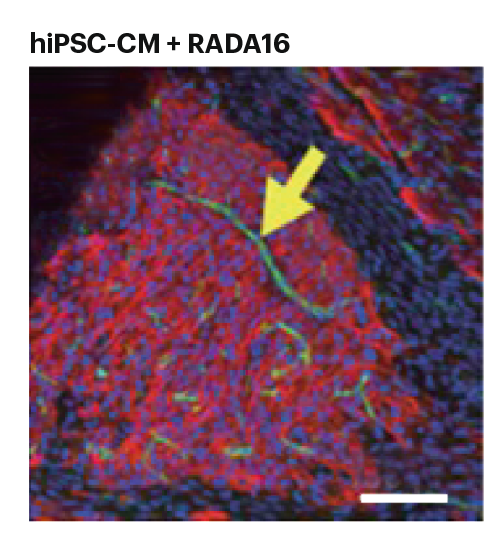
Human induced pluripotent stem cell-derived cardiomyocytes (hiPSC-CMs) are a promising platform to treat cardiac pathologies, namely infarcted tissue. Current methods for the transplantation of hiPSC-CMs into the heart often result in differentiated cells that do not fully mimic the phenotype, properties and functions of developed cardiac cells. Thus, to realize the clinical potential of hiPSC-CMs, safe and effective transplantation strategies are needed. Now, writing in Science, Richard T. Lee, Jia Liu and colleagues (Science 390, eadw4612; 2025) report injectable, self-assembling peptides that create a suitable microenvironment for the differentiation of transplanted hiPSC-CMs into the rat heart and whose functionality is measured using a bioelectronic device.
A common issue with hiPSC-CM transplantation is the spontaneous arrhythmogenic automaticity of the implanted tissue, which can result in deadly complications. Lee and colleagues design a flexible biooelectronic mesh with 32 channels that can obtain electrophysiological recordings with high spatiotemporal resolution from hiPSC-CM and native cardiac tissue. Sinus rhythms are measured for the hiPSC-CMs alone and hiPSC-CMs with RADA16 injection sites. Whereas the hiPSC-CMs alone sites generate arrhythmogenic automaticity rhythms, these undesired rhythms are not detected for the hiPSC-CMs with RADA16 sites. Longitudinal electrophysiology studies show that integration of hiPSC-CMs with RADA16 promotes a faster reduction of the percentage of arrhythmogenic automaticity compared with hiPSC-CMs. Moreover, at 135 days post-transplantation, the electrophysiology of hiPSC-CMs with RADA16 sites resembles that of the reference site in the rat heart.
This is a preview of subscription content, access via your institution